| 图片: | |
|---|---|
| 名称: | |
| 描述: | |
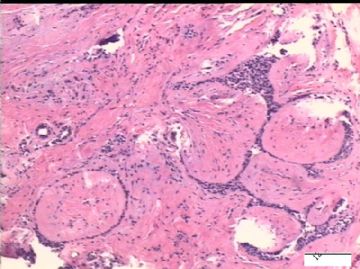
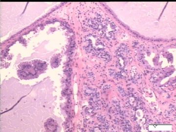
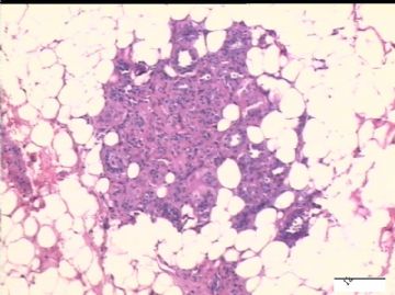
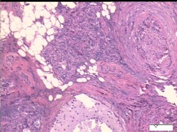
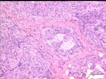
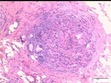
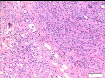
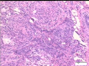
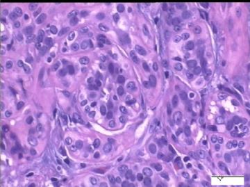
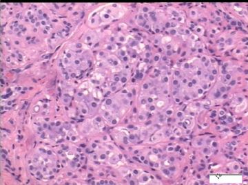
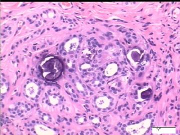
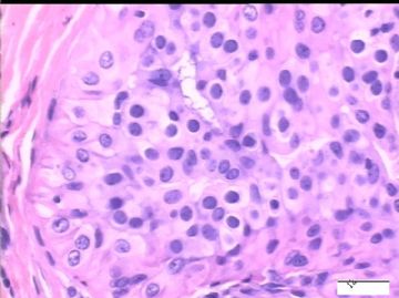
- B2431乳腺病局灶癌变?
| 姓 名: | ××× | 性别: | 女性 | 年龄: | 43岁 |
| 标本名称: | |||||
| 简要病史: | 体检偶然发现右乳腺肿块, 钼靶示不规则钙化。 | ||||
| 肉眼检查: | 大体组织 4.5 x 3.2 x 2.8 cm, 灰红色与淡黄色相间,颗粒状,无坏死。 | ||||

- xljin8
相关帖子
-
2009xiaocao 离线
- 帖子:77
- 粉蓝豆:275
- 经验:218
- 注册时间:2009-12-01
- 加关注 | 发消息
Interesting case. Thank for showing complete stains and your final diagnosis.
Most adenomyoepitheliomas (AME) are varants of introductal papilloma and small cases may arise from a lobular proliferaton or adenosis. Almost all AMEs are benign and treated with complete local excision. malignant transformation or cases with distant metastases have been described, but they are rare. The maliganancy may be limited to either epithelial or myoepithelial component or both. Evidence of malignancy includes mitotic activity, marked atypia, necrosis, invasive edges.
Above case is a difficult one. It may be AME by reviewing photos again. I am not sure the meaning of neuroinvasion. Neuroinvasion may be one of features of malignant tranformation in AMEs. I did not notice in literature. It may be a good case report especially if you have several years of follow up results.
| 以下是引用cqzhao在2010-1-10 22:02:00的发言: Interesting case. Thank for showing complete stains and your final diagnosis. Most adenomyoepitheliomas (AME) are varants of introductal papilloma and small cases may arise from a lobular proliferaton or adenosis. Almost all AMEs are benign and treated with complete local excision. malignant transformation or cases with distant metastases have been described, but they are rare. The maliganancy may be limited to either epithelial or myoepithelial component or both. Evidence of malignancy includes mitotic activity, marked atypia, necrosis, invasive edges. Above case is a difficult one. It may be AME by reviewing photos again. I am not sure the meaning of neuroinvasion. Neuroinvasion may be one of features of malignant tranformation in AMEs. I did not notice in literature. It may be a good case report especially if you have several years of follow up results. 非常感谢 Dr. Zhao 对此病例的点评。乳腺腺肌上皮肿瘤比较少见,有时生物学行为难以判断,许多问题有待研究。建议Dr. Zhao组织一个专题讨论,有兴趣的同道共同参与,提供病例。充分利用我国病例多的优势,在乳腺病理的某些方面有所建树。像日本和欧洲医生那样, 也能在世界外科病理领域争得一些话语权! 问题: 1)您建议的随访是个非常重要的判别肿瘤性质的证据。但是,如果局部癌变组织已经被完全切除,不可能再转移,5年后病人无转移,情况良好,能否定恶性诊断吗? 2)本例浸润神经的肿瘤细胞p53+是否对判断良恶性也有帮助? 谢谢! |

- xljin8
非常感谢 Dr. Zhao 对此病例的点评。乳腺腺肌上皮肿瘤比较少见,有时生物学行为难以判断,许多问题有待研究。建议Dr. Zhao组织一个专题讨论,有兴趣的同道共同参与,提供病例。充分利用我国病例多的优势,在乳腺病理的某些方面有所建树。像日本和欧洲医生那样, 也能在世界外科病理领域争得一些话语权!
Agree. I occasionally attended the lectures of Chinese pathology experts here. I always heard somethings like 国外的研究,国外的报道,国外的标准........
There are so many pathologists, so many samples. We rarely see that the pathologists in the world observe some diagnostic standards made by pathologists in China. Wish we 在乳腺病理的某些方面有所建树.
您建议的随访是个非常重要的判别肿瘤性质的证据。但是,如果局部癌变组织已经被完全切除,不可能再转移,5年后病人无转移,情况良好,能否定恶性诊断吗?
2)本例浸润神经的肿瘤细胞p53+是否对判断良恶性也有帮助?
Do not know the answer for above.
I agree.
In fact the most useful study for practice pathologists are:
have a large number of cases with histology analysis or some pathologic features, diagnostic criteria, and clinical follow up results.
You can find the most of important pathology papers published in Am J of surg path with the similar ways.
If you have several cases 乳腺腺肌上皮肿瘤 with neuroinvasion you can summary clinial, pathologic and ihc features. It should be a good paper with new findings.

