| 图片: | |
|---|---|
| 名称: | |
| 描述: | |
- B2312乳腺恶性肿瘤,请教类型?再次传图,有诊断结果了!
| 姓 名: | ××× | 性别: | 女性 | 年龄: | 56 |
| 标本名称: | |||||
| 简要病史: | 1年, | ||||
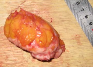
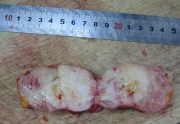
| 肉眼检查: | 如图 | ||||
-
本帖最后由 于 2010-01-16 12:37:00 编辑

- 三人行,必有我师焉,择其善者而从之,择其不善者而改之。
相关帖子
- • 左乳腺肿块
- • 乳腺肿物2---诊断?
- • 乳腺化生癌?
- • 06年的一例乳腺癌,什么类型?
- • 乳腺肿物
- • 化生性癌?
- • 女 60岁 乳腺6*7大小 切面半囊半实 红褐色
- • 可否诊断基底细胞样乳腺癌?
- • 右乳腺肿块
- • 右乳肿块,新加免疫组化结果
| 以下是引用cqzhao在2010-1-17 19:54:00的发言:
Sorry, I cannot agree with above. ER/PR/Her2 are very important factors for prognosis and chioce of treatment. We, pathologists cannot say 因为考虑到这类肿瘤ER/PR/her2一般情况下都是阴性,所以没有做. 因为ER/PR/Her2 stains were not performed,所以you cannot say they are 阴性.
|

- 三人行,必有我师焉,择其善者而从之,择其不善者而改之。
| 以下是引用向您学习在2010-1-17 19:44:00的发言:
|
Sorry, I cannot agree with above. ER/PR/Her2 are very important factors for prognosis and chioce of treatment.
We, pathologists cannot say 因为考虑到这类肿瘤ER/PR/her2一般情况下都是阴性,所以没有做.
因为ER/PR/Her2 stains were not performed,所以you cannot say they are 阴性.
| 以下是引用cqzhao在2010-1-17 0:23:00的发言:
Thank 向您学习 to ley us know your IHC results and final diagnosis. This is the way to show a case in the web. Do you know the results of ER/PR?her2? Suppose all breast carcinomas should have these stains? I am not surprised if all of them are negative. |

- 三人行,必有我师焉,择其善者而从之,择其不善者而改之。
-
本帖最后由 于 2010-01-05 11:17:00 编辑
重新发图并谈一下我的一些浅显认识,请大家指教
肿瘤切面实性,大部分为瓷白色,少部分为灰白淡红色。瓷白色区域质硬,有粘液渗出及钙化;灰白淡红色区域质软易切。
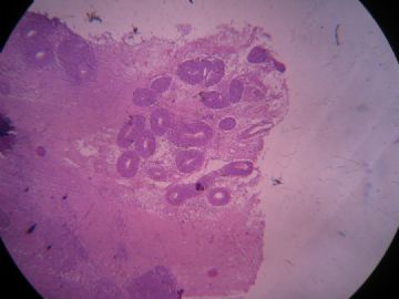
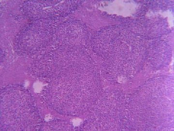
本例取材(灰白淡红色区域)主要显示的结构就像图一所示:低倍镜下看肿瘤细胞呈“巢”分布,但中、高倍镜下看(图二、三)瘤细胞“巢”周围都是坏死组织。仔细查看每一张切片发现(图四)这里邻近肿瘤边缘的地方肿瘤细胞呈弥漫分布,并且在肿瘤组织的边缘(图五)见到了因坏死被孤立出去的肿瘤细胞“巢”,在这里我想到了海洋中的岛屿。所以因为坏死而在低倍下看到一个假象:“巢”状结构(图六)。所以,我认为,每个“巢”中的血管(图七)应该就没有了诊断上的意义。
在这里(图八、九)我发现这部分肿瘤细胞胞浆透明、边界清楚。肿瘤的边缘(图十)发现有淋巴细胞浸润,在其附近(图十一)发现坏死有淋巴细胞、浆细胞的参与。图十二、十三是不是肿瘤细胞与间质细胞的移行?这些所见是否对诊断有帮助?
图十四、十五、十六为瓷白色区域

- 三人行,必有我师焉,择其善者而从之,择其不善者而改之。
-
本帖最后由 于 2010-01-01 23:59:00 编辑
Intersting growth pattern.
Suggest stains:
CK (cam 5.2, ck5/6, ae1/ae3)
Vimentin
Neuroendocrine markers: chromogranin, synaptophysin.
First we should know it is a carcinoma, sarcoma, or neuroendocrine tumor.
Thank Dr 向您学习 to share the case and hope you can continue to show us the stain result or your diagnostic result.
We should 向您学习 . Love your net name.
有趣的生长方式。
建议染色:
CK (cam 5.2, ck5/6, ae1/ae3)
Vimentin
神经内分泌标记物:CgA,Syn
首先我们要区分它是癌、肉瘤还是神经内分泌肿瘤。
谢谢“向您学习”分享病例,并希望您继续提供染色结果或者诊断结果。
我们应该向您学习,喜欢您的网名。
__abin译


