
 - WHO grade III meningeal hemangiopericytoma图1")
 - WHO grade III meningeal hemangiopericytoma图2")
 - WHO grade III meningeal hemangiopericytoma图3")
 - WHO grade III meningeal hemangiopericytoma图4")


| 图片: | |
|---|---|
| 名称: | |
| 描述: | |
- NP (2) - WHO grade III meningeal hemangiopericytoma
-
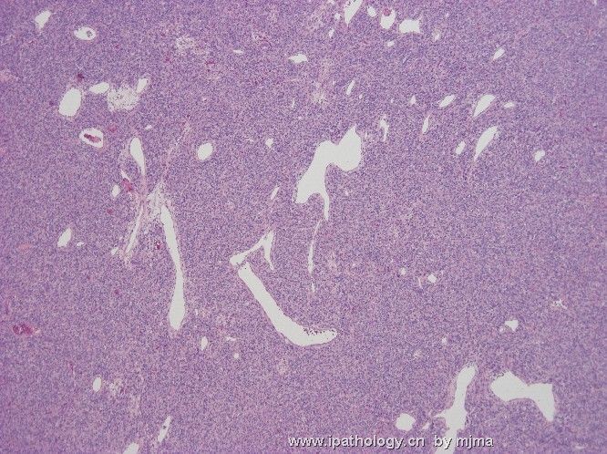
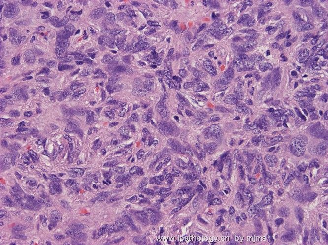
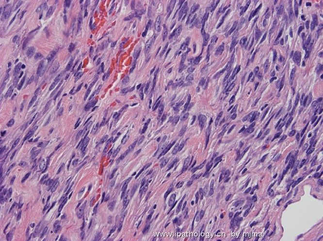
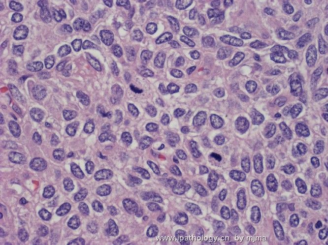
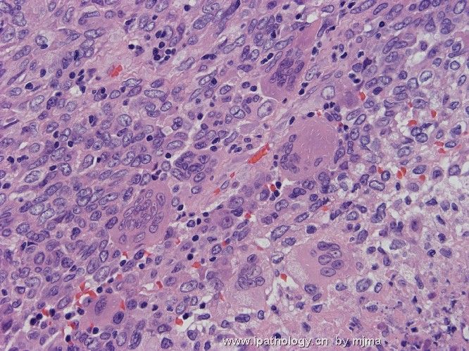
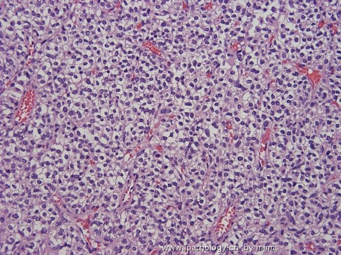
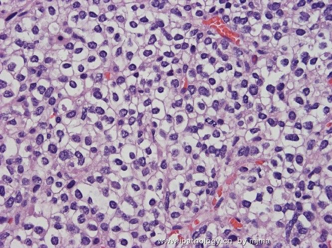
The following 4 photos were taken from intraoperative smear and frozen section of an 8 cm dura-based brain tumor in a 50-year-old woman. The surgeon noted that the tumor bled profusely during surgery. What are your differential diagnoses? Photos of paraffin sections will be uploaded in a few days.
 - WHO grade III meningeal hemangiopericytoma图1") 图1
图1 - WHO grade III meningeal hemangiopericytoma图2") 图2
图2 - WHO grade III meningeal hemangiopericytoma图3") 图3
图3 - WHO grade III meningeal hemangiopericytoma图4") 图4
图4
标签:
-
本帖最后由 于 2006-10-23 21:11:00 编辑

聞道有先後,術業有專攻
×参考诊断
-
本帖最后由 于 2006-10-23 21:21:00 编辑
| 以下是引用xiaohl 在2006-10-23 11:37:00的发言: 关于脑膜血管周细胞瘤的分级问题,我查了相关书籍和文献,一般都认为它是一低度恶性或具有恶性潜能的肿瘤,WHO2-3级.如果核异型性明显,核分裂象活跃(>5/10hpf),出现坏死和出血,那么可定为3级. |
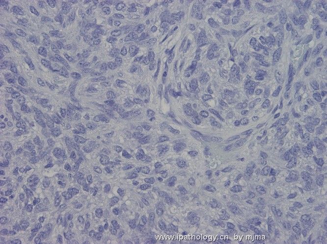
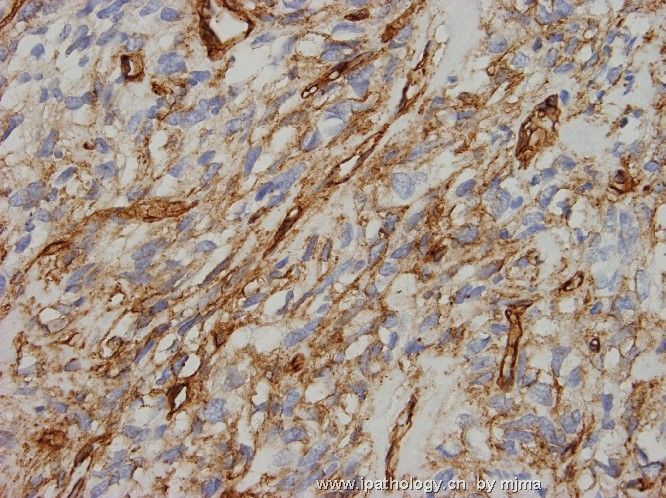
One important thing about CD34 immunostaining - classic hemangiopericytomas and fibrous meningiomas show patchy staining, whereas solitary fibrous tumors show diffuse and intense staining. This particular case shows only patchy and not diffuse staining - a point that may comes in handy when distinguishing a solitary fibrous tumor from a low-grade hemangiopericytoma.
聞道有先後,術業有專攻
-
本帖最后由 于 2006-10-23 21:12:00 编辑
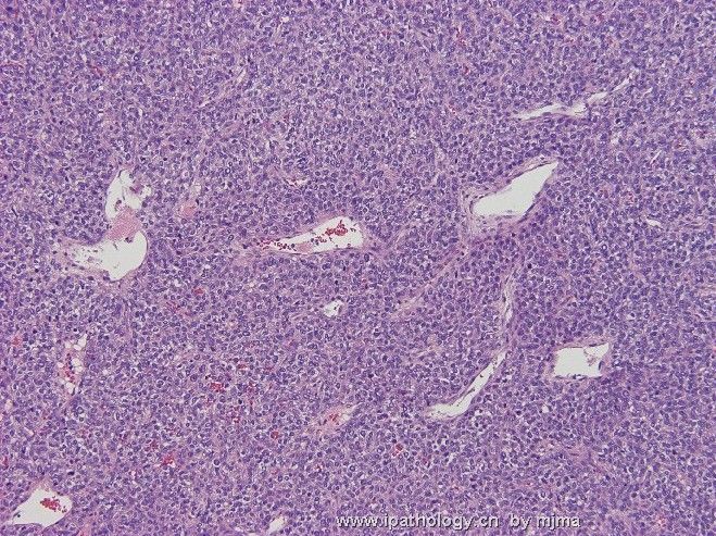
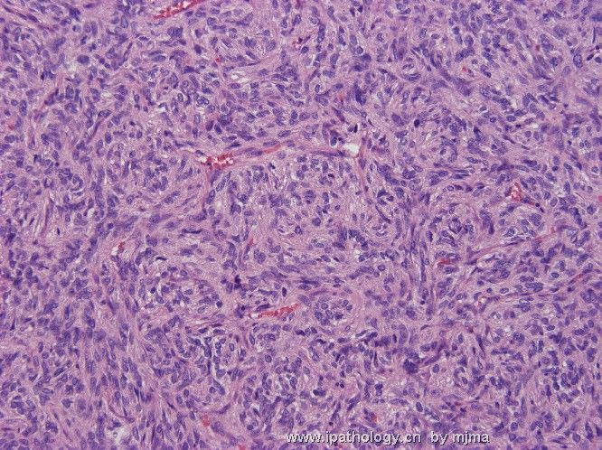
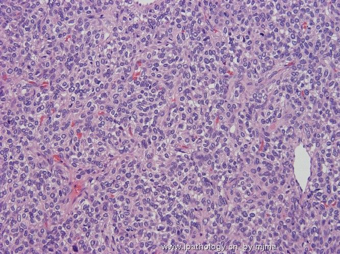
This is a case of high grade (WHO grade III) meningeal hemangiopericytoma. All hemangiopericytomas are malignant. Using a few histologic features like mitotic counts and necrosis, hemangiopericytomas (in CNS or elsewhere) can be graded as high (WHO grade III) or low grade (WHO grade II).
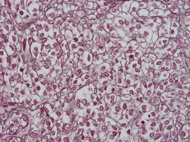
Figures 1~5 of my second batch of photos display classic H&E features of hemangiopericytoma, and pericellular reticulin fibrils are quite characteristic (Figure 13). In addition, I wish to illustrate the less publicized histologic features of hemangiopericytomas. All photos are taken from the same tumor. Figure 6 really mimics solitary fibrous tumor. Figure 7 is from an area of hyalinized fibrosis, often seen in central areas of the tumor. Figures 11 and 12 show clear cells that may be mistaken for oligodendroglioma. Figure 8 shows abundant mitotic figures in this case. Figure 9 shows tumor necrosis, and Figure 10 displays multinucleated osteoclast-like giant cells that may be present with or without associated necrosis.
Ever since solitary fibrous tumor became a popular disgnosis about 10 years ago, the existence of meningeal hemangiopericytoma has been questioned. I have never lost faith in its existence in the CNS. Meningeal hemangiopericytomas are notoriously vascular and bleed readily during surgery. They may seed the neuraxis via CSF, recur locally after resection, and metastasize to extraneural sites. Therefore, post-operative irradiation is needed.
聞道有先後,術業有專攻
-
本帖最后由 于 2006-10-17 10:08:00 编辑
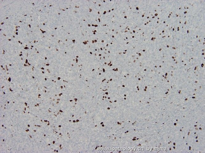
These are photos taken from HE stain (1~12), reticulin silver stain (13), EMA (14), CD34 (15) and MIB-1 (16) immunostains. What is your diagnosis? How do you grade this lesion according to WHO 2000?
 图1
图1 图2
图2 图3
图3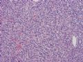 图4
图4 图5
图5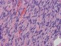 图6
图6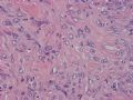 图7
图7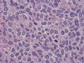 图8
图8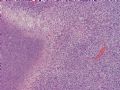 图9
图9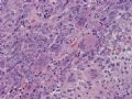 图10
图10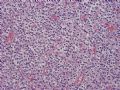 图11
图11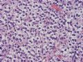 图12
图12 图13
图13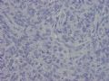 图14
图14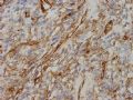 图15
图15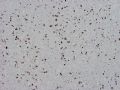 图16
图16
聞道有先後,術業有專攻


