| 图片: | |
|---|---|
| 名称: | |
| 描述: | |
- A pap case yesterday-the patient will have biopsy soon
-
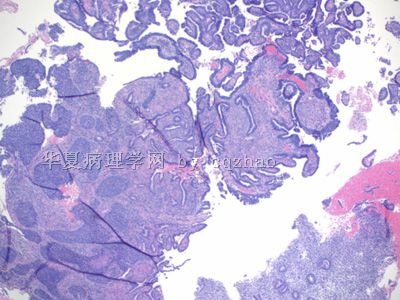
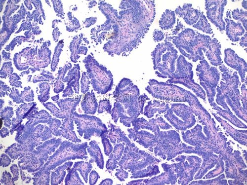
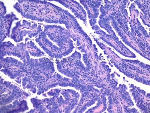
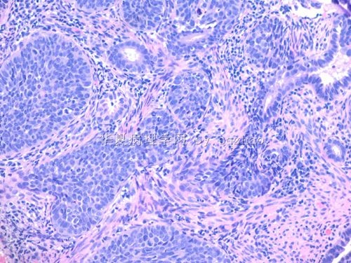
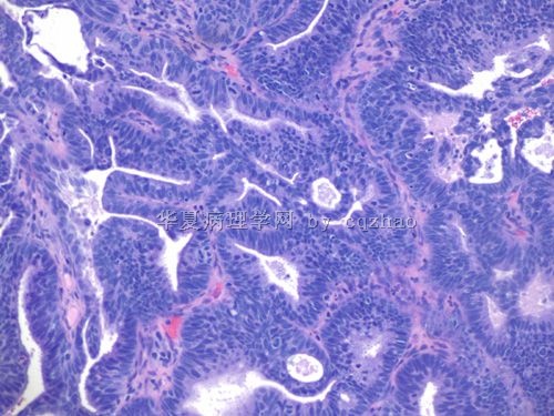
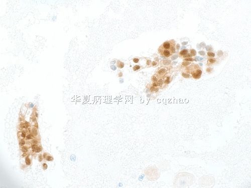
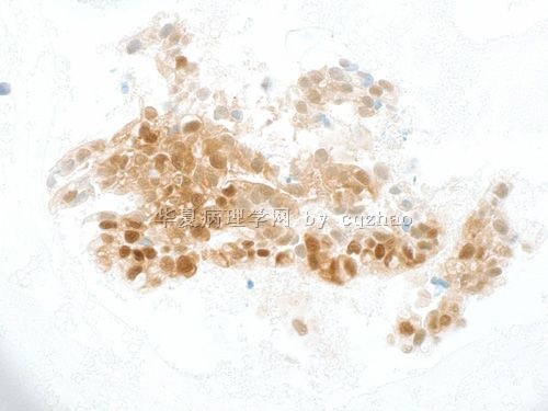
I just got the p63 and ck5/6 stains for the biopsy specimen in floor 28. Are the solid areas poorly differentiated adenocarcinoma with solid growth or squamous cell carcinoma component? Guess what? My original impression is wrong. See above. I will take some photos for your own judgment. I feel this case becomes interesting now.
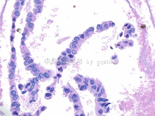
For the histology specimen above we call invasive adenocarcinoma, endometrioid type. The primary can be endocervical or endometrial.
Base on patient's age, Pap smear, and histolgy I personally think it will be an endocervical ca as 青青子矜 mentioned before.
The solid part in the biopsy specimen may be squamous component or adenocarcinoma with solid growth pattern (filling entire glands). I favor it is adenocarcinoma component also. Any way I may order some IHC stains (p63, ck5/6) for education purpose (will not charge the patient).
I will let you know the result when I have it.
-
本帖最后由 于 2009-09-23 23:48:00 编辑
| 以下是引用zxh887119在2009-9-22 21:55:00的发言:
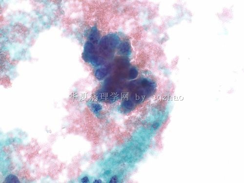
这是一个非常精彩的病例,尤其是腺细胞的病变,如果从中能学到什么,那将是我们的荣幸。临结尾之时,我提个问题,望不吝赐教。 青青子矜 和掌心0164 在看图二时有分歧(对第二图的分歧在第6和7楼)。 青青子矜 :“图2给我太强烈的羽毛感” 掌心0164 :“但是图2我感觉不像腺,没有那种像羽毛杨的核垂直于细胞团长轴;反而像累腺的鳞状上皮病变。” 最后的结果已经出来了,回过头来看,我们怎么来理解图二呢?顺便谈谈当我们拿起这张液基片时看到这几个细胞的诊断思路及回过头来的总结。 谢谢! |
青青子矜 and 掌心0164 will give you good answers.
It is very important for cytopathologists to check the histologic follow up results. Review of cytology-histology, histology-cytology, over and over. It is the most important way to learn cytology.
Ok, I need to work on my gyn biopsy for my 54 cases today. Luck part is that i have an excellent gynecologic pathology-fellow to work with me this week.
对照随后的组织学结果对细胞病理医生很重要,不断回顾细胞--组织、组织--细胞,循环往复,这是学习细胞学的最重要方法。
好,我要去处理今天的54例妇科活检了,幸运的是本周我有个能干gynecologic pathology fellow (one year gynecologic fellow after 4 or 5 year pathology resident traning)。
-
本帖最后由 于 2009-09-21 12:39:00 编辑
Now there is no problem it is a carcinoma. (现在,这是一个癌没有疑问)
The questions are (问题如下:)
1. carcinoma types (1、癌的类型)
a. endometrial with focal squamous differentiation or adenosquamoys ca like 掌心0164 mentioned above.(a、内膜来源伴有局灶性鳞状分化或如掌心0164提到的腺鳞癌)
b. endometrial with focal solid growth(b、内膜来源伴有实性生长方式)(按青青姐姐指点做了修改)
c. serous carcinoma(c、浆液性癌)
2. primary: endocervical vs. endometrial(2、原发部位:颈管或内膜)
My colleaque took care of the biopsy specimen and I cosigned the case. She did not do IHC for the biopsy specimen to work-up because she discussed with gynecologist who would do hysterectomy for this patient. (我同事负责这个活检标本和我一起签发了这个报告。她没用活检标本做免疫组化,因为她已经跟妇科医生讨论这个病人要进行子宫切除)
Anyway you can try to answer my above questions based on the photos if you are interested. I will paste the hysterectomy specimen result when I have. (还有,如果您感兴趣,基于那些照片试着回答我上面的问题。等我有了子宫切除之后的结果,我会贴在这里)-
本帖最后由 于 2009-09-20 09:18:00 编辑
| 以下是引用掌心0164在2009-9-19 8:50:00的发言:
谢谢赵老师精彩的病案;这个组织学:(宫内膜)腺鳞癌;回头看细胞学;赵老师叫的AGC-N和ASC-H是最为准确和合适的报法了。赵老师您这个病案做没有做宫颈活检呢?宫颈和宫颈管情况怎么样呢? |
-
本帖最后由 于 2009-09-19 19:05:00 编辑
I did some IHC for the cell block. Considering the cost-effect, it is not necessary to do IHC now because patient will have biopsy soon. IHC can be do for biopsy specimen if needed.
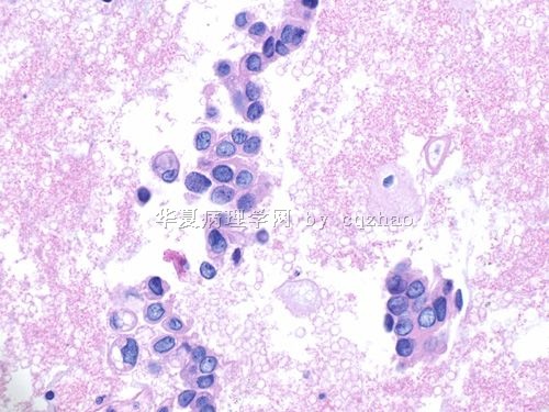
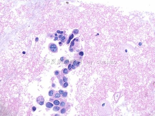
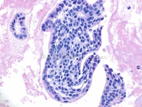
The glandular cells in cell block are negative for p53, WT1, and ER, and positive for p16. I did these IHC and try to know the carcinoma types (serous, endometrioid) and origins (endocervical or endometrial).(我用细胞腊快做了一些免疫组化。因为病人很快会有活检;考虑到成本,免疫组化不是必须的,如果确实需要我们会用活检标本再做。在细胞腊块中的腺细胞p53, WT1, 和ER都是阴性;而P16阳性。我做这些免疫组化想知道癌症类型<浆液性,内膜来源>和原发部位<颈管或内膜>)
名称:图1
描述:图1
名称:图2
描述:图2
-
本帖最后由 于 2009-09-19 18:53:00 编辑
| 以下是引用Danrs在2009-9-15 21:34:00的发言:
细胞学,核大深染,我觉得还是恶性的可能大。到底是鳞状上皮来源还是腺体来的,似乎更像腺体。 后面的切片,不知道是怎么来的,似乎只有腺体,看不到间质。如果是宫颈脱落细胞的离心沉淀包埋切片,腺体居然还能保存这么好。腺体不像正常的宫颈腺上皮,但是个别细胞也像粘液腺上皮。考虑宫颈来源的。 |
-
本帖最后由 于 2009-09-19 18:42:00 编辑
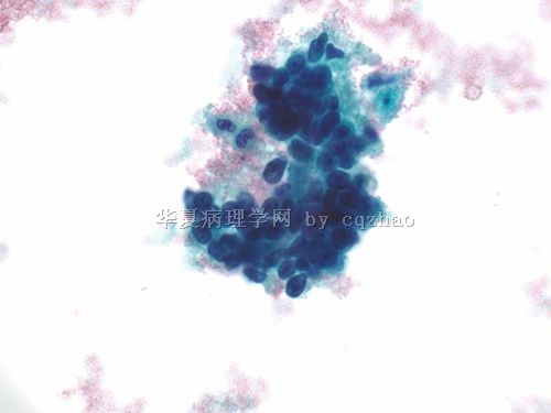
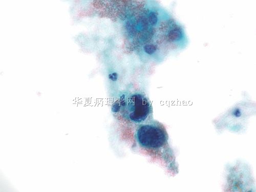
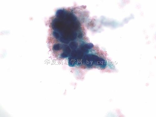
I think most of you had very reasonable interpretation based on the Pap photos. Bottomline is the women should have tissue sampling. It is too undercall if we call ascus for these kinds atypical cells within the backgrouding of blood.
Friday I signed out this case as AGC-favor neoplasm and ASC-H. The clusters of cells are more like glandular lesion. Two single cells and the second cluster show some squamous features as 掌心0164 mentioned. Anyway Friday I discussed the case with primary doctor and told him that I thought it was adenocarcinoma case and patient should have endometrial and endocervial sampling even I released the case with AGC-N and ASC-H. However I am not sure the origin, endometrial or endocervical, even though it may be a cervical orgin based on the age of 45. (基于照片我认为大家绝大部分人的解释是合理的;底线是这个病人必须做组织活检。在这种血性背景下这些类型的非典型细胞,如果我们叫ASCUS是不够的。礼拜五,我签发这个病例为AGC-FN和ASC-H。那些成团的细胞更像腺上皮的病变。两个单个细胞和第二图的细胞团显示了一些鳞状细胞的特征。还有礼拜五我跟我的住院医生讨论这个病案的时候;即使我报的是AGC-FN和ASC-H,但是我认为是一个腺癌病例必须有内膜和颈管的活检。即使由于45岁的年龄可能来源宫颈;但是我不能肯定原发部位:内膜或颈管)掌心0164翻译
-
本帖最后由 于 2009-09-19 18:22:00 编辑
I asked the lab to make a cell block by using the residual Pap liquid and got it today. Paste here a few cell block photos with high power. (我让实验室技术员用剩下的液体做了细胞腊块,今天看到了切片;以下是一些细胞腊块的高倍照片)
名称:图1
描述:图1
名称:图2
描述:图2
名称:图3
描述:图3
名称:图4
描述:图4
