| 图片: | |
|---|---|
| 名称: | |
| 描述: | |
- A pap case yesterday-the patient will have biopsy soon
Continue:
7. add 3-4 drops of thrombin and mix gently, swirling the tube.
8. allow to stand 1-2 min. If a clot does not form, use a few additional drops of plasma.
9.pull clot onto lens paper premoistened with saline solution and work ito an even ball of materials.
10. fold paper over clot carefully.
11. place clot in cassette and place in formalin container.
12. than treat it like biopsy specimen.
I think it will be similar for this procedure among all cytology lab. Do not ask me the detailed question. I never did it in person. Above is our protocal.
Hope it can help. cz
| 以下是引用轻羽飞扬在2009-9-27 11:17:00的发言: 赵老师有时间的话能否介绍一下你们细胞块的制作过程。谢谢! |
Remember the home work. Today I asked people in Cyto lab to get a protocal.
We use ThinPrep Pap. It is 20 cc fluid. 4-6 cc for a Pap depending on the cell concentrition). If the patient did not do the HC2 hpv test, 4 cc fluid will be take in case use for hpv test. Then
1 pour all specimen into 50 ml centrifuge tube and centrifuge for 5 m.
2. pure supernatant into original container.
3. add 5 cc of saline solution to pellet and swirl gently to mix specimen (but do not vortex)
4. centrifuge for 5 min
5. pour or pipette off saline
6. add 3-4 drops of room temperature pooled plasma and mix by gently swirling the tube
sorry, I need to leave here now
-
本帖最后由 于 2009-10-18 10:00:00 编辑
Pts with stage II should get radiation and chemotherapy. This is why we may not see the hysterectomy specimen for this patient.
you can check detailed information for staging and treatment for cancer from NCCN guideline -the version of translation, provided by Dr. Lotus from another topic.
临床II期以上的病人应该是放化疗,这是为何我们看不到这个病人的子宫切除标本的原因。
你们可以参照NCCN指引(website provided by Dr. Lotus )来核对肿瘤临床分期和治疗的详细资料。详见下列网址:
另外:我记得应该是具体到IIb以上的病人才失去手术机会,只做放化疗。(青青子矜)
-
本帖最后由 于 2009-10-18 09:53:00 编辑
- Stage IA: Invasive cancer identified only microscopically. Invasion is limited to measured stromal invasion with a maximum depth of 5 mm and no wider than 7 mm.
- Stage IA1: Stage IA1: Measured invasion of the stroma no greater than 3 mm in depth and no wider than 7 mm diameter.
- Stage IA2: Stage IA2: Measured invasion of stroma greater than 3 mm but no greater than 5 mm in depth and no wider than 7 mm in diameter.
- Stage IB: Stage IB: Clinical lesions confined to the cervix or preclinical lesions greater than Stage IA. All gross lesions even with superficial invasion are Stage IB cancers.
- Stage IB1: Stage IB1: Clinical lesions no greater than 4 cm in size.
- Stage IB2: Stage IB2: Clinical lesions greater than 4 cm in size.
- Stage IIA: No obvious parametrial involvement. Involvement of up to the upper two-thirds of the vagina.
- Stage IIB: Obvious parametrial involvement, but not into the pelvic sidewall.
- Stage IIIA: No extension into the pelvic sidewall but involvement of the lower third of the vagina.
- Stage IIIB: Extension into the pelvic sidewall or hydronephrosis or non-functioning kidney.
- Stage IVA: Spread of the tumour into adjacent pelvic organs.
- Stage IVB: Spread to distant organs.
FIGO stage: Tomor is metastatic to the wall of bladder. It should be at least stage IIB. If tumor metastatic to the mucosa of bladder and recturm or beyond true pelvis it is stage IV. We do not know if the tumor speads to the low third of vagina or pelvic wall. Bladder wall is a parametrial organs. This is why I think the tumor is at least stage IIB. Correct me if i am wrong.
FIGO分期:肿瘤转移到膀胱壁,应该是至少临床IIB以上。如果肿瘤转移到膀胱粘膜层和rectum(这里好像有笔误,我的理解是rectum--直肠--((I am wrong, you are right)或真骨盆,应该是临床IV期。我们不知道如果肿瘤超出了阴道的下1/3或骨盆壁,其分期又是怎样?膀胱壁是子宫旁器官,这是我认为该肿瘤的临床分期至少在IIB以上的原因。如果我错了,请纠正。
呵呵,赵老师谦虚。(青青子矜)
具体FIGO分期不再翻译,请感兴趣的网友自己参考中文译本
|
FIGO STAGING OF CERVICAL CARCINOMAS | |
| Stage I | |
| Stage I is carcinoma strictly confined to the cervix; extension to the uterine corpus should be disregarded. The diagnosis of both Stages IA1 and IA2 should be based on microscopic examination of removed tissue, preferably a cone, which must include the entire lesion. |
|
| Stage II | |
| Stage II is carcinoma that extends beyond the cervix, but does not extend into the pelvic wall. The carcinoma involves the vagina, but not as far as the lower third. |
|
| Stage III | |
| Stage III is carcinoma that has extended into the pelvic sidewall. On rectal examination, there is no cancer-free space between the tumour and the pelvic sidewall. The tumour involves the lower third of the vagina. All cases with hydronephrosis or a non-functioning kidney are Stage III cancers. |
|
| Stage IV | |
| Stage IV is carcinoma that has extended beyond the true pelvis or has clinically involved the mucosa of the bladder and/or rectum. |
-
本帖最后由 于 2009-10-18 09:18:00 编辑
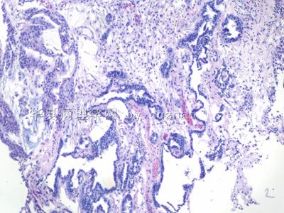
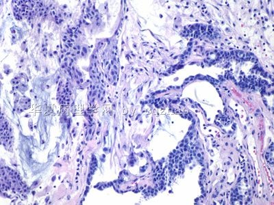
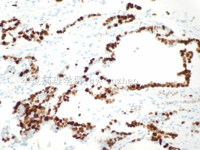
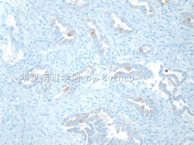
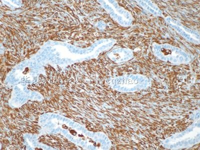
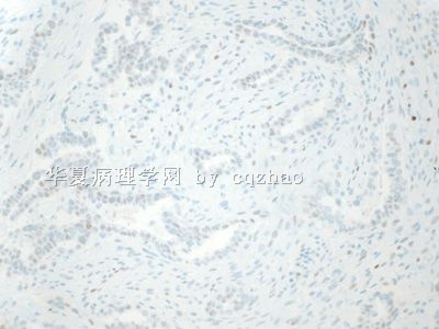
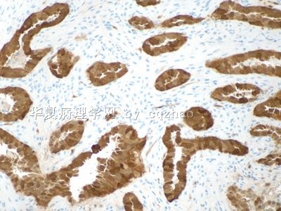
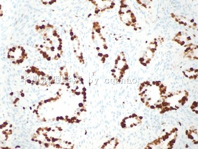
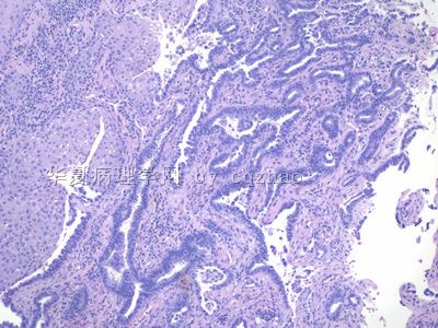
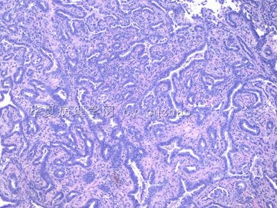
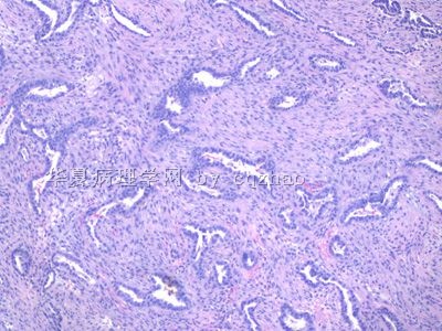
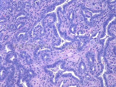
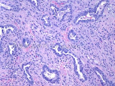
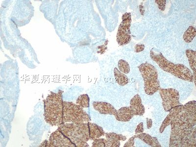
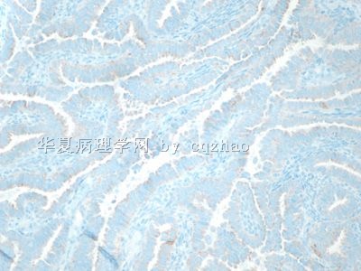
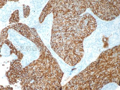
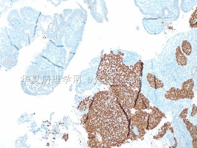
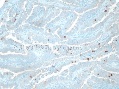
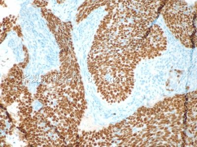
adenocarcinoma component of this case is positive for p16, CEA, negative for vimentin and ER. So it is very calssic pattern for endocervical adenocarcinoma.
Tis is a very classic invasive adenosquamous carcinoma of cervix.
本例的腺癌成分是p16、 CEA阳性,vimentin 、ER阴性,所以这是个经典的宫颈腺癌表达模式。
这是例典型的宫颈浸润性腺鳞癌。(青青子矜译)
| 以下是引用青青子矜在2009-10-15 18:13:00的发言:
懒了很多天,今天终于得空重新看完了全帖,现在来回答这些问题。 1、宫颈腺癌。“肿瘤细胞中p16阳性,CEA、Vimentin和ER为阴性"但一般宫颈腺癌的CEA也常常是阳性,呵呵; 2、宫颈腺鳞癌(有明确腺癌成分,另实性巢团部分CK5/6和P63都为阳性,这是鳞的标记),转移或浸润到膀胱壁; 3、不需要了。放化疗时首选; 4、膀胱壁有转移,至少是临床III期以上(盆腔内),四期是有远处转移。而宫颈癌IIb以上就不再是外科手术切除的指征,鳞癌对放疗敏感,但此例的预后就很难说了。 期待赵老师指正。感谢海浪的不辞劳苦。 |
| 以下是引用cqzhao在2009-9-28 4:14:00的发言:
Hope some ones can answer the following questions. 1. Tumor cells are positive for p16, cea, negative for vimentin and ER. They are very typical pattern for ( ? ) adenocarcinoma. 2. Based on the H&E, IHC, clinical prsentation, what is the full dx for this case? 3. Do you think women should have hysterectomy? Thank all of you reading or participating discussion for this topic. Basically I finish my duty for this topic. cz 尝试翻译:(海浪信使) 希望(有兴趣的)一些网友可以回答下列问题。
1、肿瘤细胞中p16阳性,CEA、Vimentin和ER为阴性。他们是非常典型的(?)腺癌模式。 2、based on HE、免疫组化、临床资料, what is your final diagnosis? 3、你认为患者应该做子宫切除术吗?
4. What is staging (at least) for this case?
谢谢各位阅读或参与这一病例的讨论。基本上我完成了这个病例我所做的工作。 Thank 海浪信使. cz
|
seems our pathologists are not interested to these clinical questions.
Clearly this is invasive adenosquamous carcinoma.
You can find answers from the web about staging and standard treatment for this patient.
http://www.nccn.org/professionals/physician_gls/f_guidelines.asp
-
本帖最后由 于 2009-09-28 11:40:00 编辑
recently I noticed Pap section is very hot due to the effort of benzhu, assistants, and all of you. It is great. Cervical cancer screening is a very important work in China. We still have a long way to 达到world advanced level. It needs all people 共同effort including our pathologists.
Suggestion: When you have histologic follow up result for the Pap smears you pasted in the web, please paste the photos of histologic results if you can. This is a good way for us to learn. Thanks, cz
尝试翻译(海浪信使):
最近我留意到宫颈细胞学版块在版主、助理和大家的共同努力下变的很火!这是非常好的。宫颈癌筛查在中国是一个非常重要的工作,仍然需要漫长的道路才能达到世界先进水平。它需要包括我们的病理学家在内的所有人的共同努力。
建议:当你在宫颈抹片的帖子中跟进报告组织病理学检查结果的时候,请附上组织病理学结果的照片,如果你能够做到的话。这是一种很好的学习方法。谢谢!
-
本帖最后由 于 2009-09-29 05:10:00 编辑
Hope some ones can answer the following questions.
1. Tumor cells are positive for p16, cea, negative for vimentin and ER. They are very typical pattern for ( ? ) adenocarcinoma.
2. Based on the H&E, IHC, clinical prsentation, what is the full dx for this case?
3. Do you think women should have hysterectomy?
Thank all of you reading or participating discussion for this topic. Basically I finish my duty for this topic. cz
尝试翻译:(海浪信使)
1、肿瘤细胞中p16阳性,CEA、Vimentin和ER为阴性。他们是非常典型的(?)腺癌模式。
2、based on HE、免疫组化、临床资料, what is your final diagnosis?
谢谢各位阅读或参与这一病例的讨论。基本上我完成了这个病例我所做的工作。
Now I think every one should know the diagnosis. 掌心0164 is a 天才. I do not know what you can be if you learn the histology. Ha, ha.
1. Please review the Pap photos again after you know the histologic diagnosis. think over the features of cytology and histology. Learing from the true case is better than learing from the text book.
2. The origin: endocervical or endometrial. Based on all the information above I think we know most likely it is a xxxx tumor. You can fill in the xxxx by yourself.
3. I showed the IHC to the primary pathologist who took case of the biopsy specimen. She wrote an addendum for the case.
This is not a retrospective review case. I am working with your guys together for this case step by step. Sometimes pathology work is interesting.
Thanks, cz


