| 图片: | |
|---|---|
| 名称: | |
| 描述: | |
- 右肾上腺肿块 帮忙会诊!(十分罕见,多发表意见呀!)
-
chengs2001 离线
- 帖子:76
- 粉蓝豆:12
- 经验:87
- 注册时间:2006-09-27
- 加关注 | 发消息
-
本帖最后由 于 2009-09-13 19:32:00 编辑
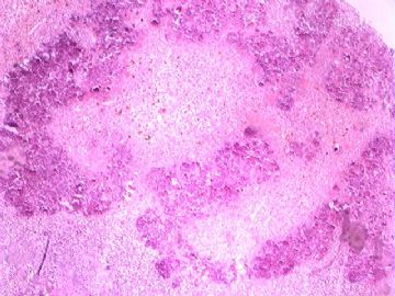
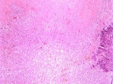
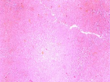
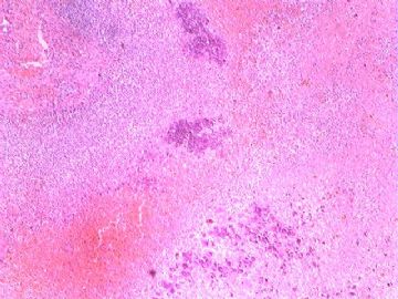
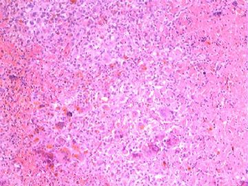
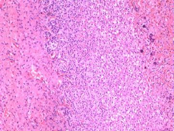
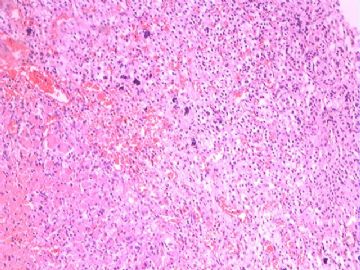
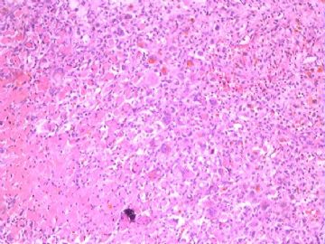
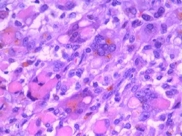
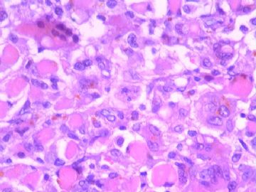
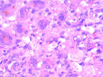
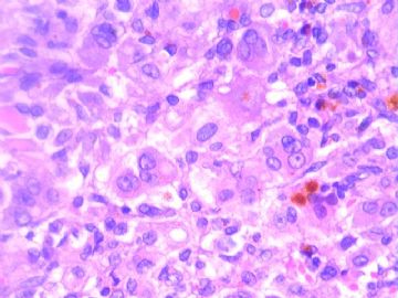
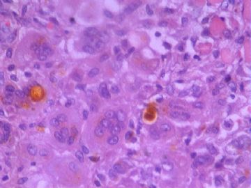
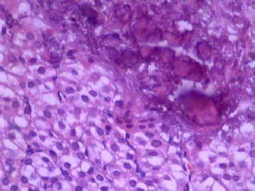
There are old hemorrhage, multinucleated giant cell reaction, calcification and recent hemorrhagic infarction in the adrenal mass containing residual adrenal cortex. I do not think this is neuroblastoma, ganglioneuroblastoma, pheochromocytoma or adrenal cortical adenoma. Figures 9 and 10 suggest nephrogenic adenoma, but the patient's young age, anatomic location and other features do not support this possibility. Like some who have commended, I am not certain this is a neoplasm. I don't think this is Rosai-Dorfman disease.
肾上腺肿物,见残留的肾上腺皮质,有陈旧出血、多核巨细胞反应、钙化和新鲜出血性梗死,我认为不是神经母细胞瘤、节细胞神经母细胞瘤、嗜铬细胞瘤或肾上腺皮质腺瘤。图9、10提示肾源性腺瘤样,但患者年幼、解剖部位和其它特征不支持这种可能性。象前面一些网友提到的,我不能确定是肿瘤性病变。也不考虑Rosai-Dorfman 病。
liguoxia71试译。

聞道有先後,術業有專攻
-
chengs2001 离线
- 帖子:76
- 粉蓝豆:12
- 经验:87
- 注册时间:2006-09-27
- 加关注 | 发消息
-
新生儿肾上腺疾病遇到的极少,手头有本Bostwick和Liang Cheng主编的第二版Urologic surgical pathology,翻了一下,可能符合的病为肾上腺cytomegaly (adrenal cytomegaly 不知中文咋翻)。先天性肾上腺cytomegaly 通常是偶然发现的(大体上可正常),被报道在3%的新生儿和6.5%的成熟流产儿尸检中发现。Cytomegaly影响胎儿(暂时性)肾上腺皮质的细胞,可以是双侧、单侧、局灶、弥漫,巨细胞有增大的、染色质深的细胞核和体积增多的胞质,核可以有显著的多形性,偶然可含核内假包涵体,尽管有显著的不正常的细胞核,但分裂像不存在。
adrenal cytomegaly是Beckwith-Wiedemann综合症的特征性成分……。主要在11P15区域内的生长调节基因失功能,导致正常生长失控,并增加某些肿瘤的发生率。 在这种疾病中,肾上腺可增大,同时重量可增至16 g。奇怪的是肾上腺的嗜铬组织可能增生或不适时的成熟了。还可能有肾和胰腺的内脏肿大,有些婴儿还发展严重的新生儿低血糖,可致死……
同时请参看一下Dr.quhong对肾上腺发育的描述。
-
liguoxia71 离线
- 帖子:4174
- 粉蓝豆:3122
- 经验:4677
- 注册时间:2007-04-01
- 加关注 | 发消息
-
liguoxia71 离线
- 帖子:4174
- 粉蓝豆:3122
- 经验:4677
- 注册时间:2007-04-01
- 加关注 | 发消息
-
肾上腺巨细胞症(anrenal cytomegaly)是一种非常罕见的先天性疾病,国内报道较少。大多数于胎儿或新生儿尸检中发现。患儿往往大体表现正常,组织学显著特征是胎性皮质细胞在细胞大小、形状及细胞结构上发生变化。细胞呈多角形,大小可达直径100μm或更大。染色质呈不规则团块,核中可见单个或多个轮廓清楚的嗜酸性球形小体。有人认为这些球形小体(假包涵体)是凹入核内的胞质。细胞质呈红染颗粒状或聚集为团块状,并可见泡沫状细胞质。这种皮质巨细胞可见于先天性巨细胞性肾上腺发育不全,死产及新生儿发生约占1%。此外它还是Beckwith-Wiedemann综合征的一部分,该综合征包括一系列先天性发育不良,即胰腺增生、低血糖、双侧肾上腺增生、脐突出、巨舌症等。

- 三人行,必有我师焉,择其善者而从之,其不善者而改之。
-
chengs2001 离线
- 帖子:76
- 粉蓝豆:12
- 经验:87
- 注册时间:2006-09-27
- 加关注 | 发消息
-
本帖最后由 于 2009-09-16 01:31:00 编辑
在读我的意见之前,要知道我不是儿科病理学者,对婴儿肾上腺组织学知识掌握的也有限。下面我引用的是Stephen Sternberg撰写的第二版病理医生专用组织学中的一段话(1110页):妊娠末期,胎儿临时皮质成分占腺体大部分。出生后几小时内,临时的皮质成分开始明显充血、变性。7-10天后,开始变得结构紊乱、坏死。周围细窄带内的细胞团存活并成为以后永久皮质的来源。看来出生后2周内胎儿皮质有坏死是可以的。所以,这个年龄,皮质部位有坏死是正常发育过程,不一定是肿瘤性坏死。前8张图可能显示了这一正常生理过程。当然,这仅是我个人观点,你们不必太当真。免疫组化结果解释困难,它标出了2种成分-T淋巴细胞和巨噬细胞,是否是坏死的主要反应细胞?CD31主要是标记内皮细胞的,是不是我们想的那样显示了血管着色?我们需要有儿科病理知识的病理学者来解释这个病例。(liguoxia试译)
Before you read my following comment, I would like you to keep this in mind. I am not a pediatric pathologist, with only limited knowledge of baby adrenal gland histology.
Here is a paragraph I quoted from "Histology for Pathologist" (2nd edition), a book written by Stephen Sternberg.
"At the end of gestation, the provisional cortex accounts for the bulk of the gland. Within hours of birth, it becomes acutely congested and starts to degenerate. At the end of 7 to 10 days, the provisional cortex is largely disorganized and necrotic. The narrow peripheral band of cell clusters survives and becomes the source of the permanent cortex" (page 1110).
It seems that the provisional cortex or fetal cortex is supposed to have necrosis in two weeks after birth. Therefore, at this age, the necrosis within the cortical location is a normal process, not necessarily a tumor necrosis. The above first 8 photos probably demonstrate this normal process. Of course, this is my personal opinion. You don't need to take it too seriously.
The immunohistochemical results are difficult to interpret. They highlight two populations of cells: T-lymphocytes and macrophages. They are the major cells reactive to necrosis, aren't they? CD31 is primarily an endothelial marker. If CD31 stains vessels, that is what we expected. We need a pathologist with pediatric pathology background to explain this case.
| 以下是引用liguoxia71在2009-9-14 11:26:00的发言: 肾上腺巨细胞症(anrenal cytomegaly)是一种非常罕见的先天性疾病,国内报道较少。大多数于胎儿或新生儿尸检中发现。患儿往往大体表现正常,组织学显著特征是胎性皮质细胞在细胞大小、形状及细胞结构上发生变化。细胞呈多角形,大小可达直径100μm或更大。染色质呈不规则团块,核中可见单个或多个轮廓清楚的嗜酸性球形小体。有人认为这些球形小体(假包涵体)是凹入核内的胞质。细胞质呈红染颗粒状或聚集为团块状,并可见泡沫状细胞质。这种皮质巨细胞可见于先天性巨细胞性肾上腺发育不全,死产及新生儿发生约占1%。此外它还是Beckwith-Wiedemann综合征的一部分,该综合征包括一系列先天性发育不良,即胰腺增生、低血糖、双侧肾上腺增生、脐突出、巨舌症等。 |
欣赏肾上腺巨细胞症(anrenal cytomegaly)这个诊断.
查过一些文献,这个诊断好!
liguoxia71医生不简单,更欣赏这个座右铭:三人行,必有我师焉,择其善者而从之,其不善者而改之。自勉.

- 王军臣

