| 图片: | |
|---|---|
| 名称: | |
| 描述: | |
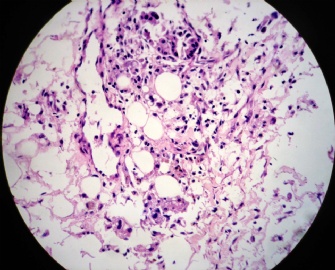
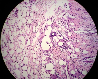
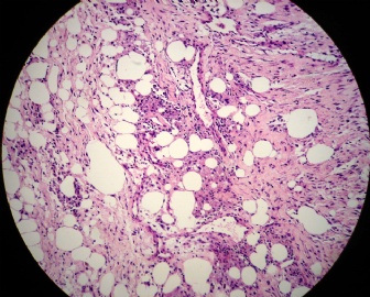
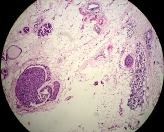
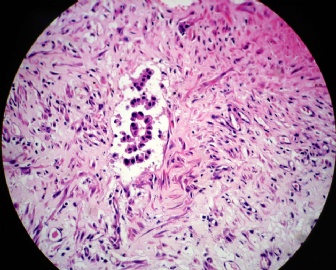
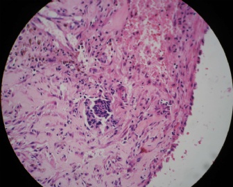
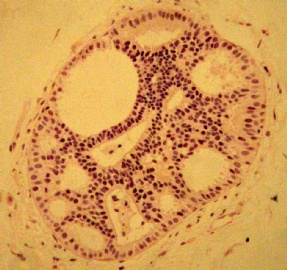
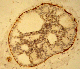
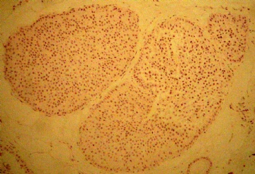
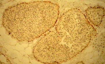
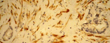
- B2154借我一双慧眼吧:有无浸润?
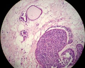
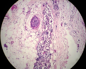
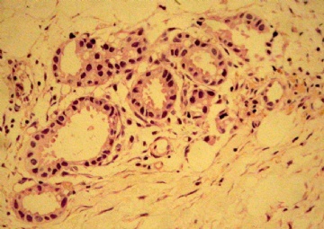
女,51岁,乳头溢血。
大体:囊内乳头状病变。
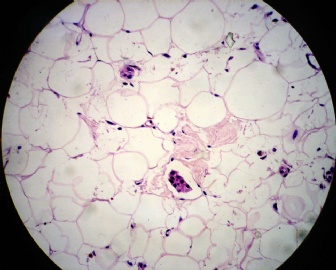
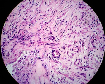
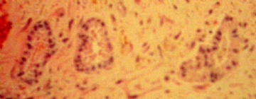
镜下:囊内乳头状肿瘤;囊壁DCIS;囊壁可疑浸润。

华夏病理/粉蓝医疗
为基层医院病理科提供全面解决方案,
努力让人人享有便捷准确可靠的病理诊断服务。
相关帖子
- • 乳腺肿瘤?
- • 右乳包块(镜下富于粘液)
- • 左乳肿块
- • 边界清楚的乳腺包块
- • 乳腺肿块,请会诊
- • 乳腺癌?
- • 左乳腺肿块,新加免疫组化
- • 左乳肿块,新加免疫组化
- • 乳腺包块-请会诊
- • 左乳肿块
-
本帖最后由 于 2009-07-21 20:00:00 编辑
谢谢Dr.Zhao,这是档案片(读片会资料),最近翻出来学习,纯粹为了学习而讨论,因此应该算学习病例。
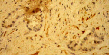
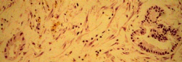
这例可以肯定的有:囊内乳头状癌,囊壁DCIS。仔细观察过HE切片和肌上皮标记,这些小腺管周围确实没有肌上皮。但是没有做基底膜标记,而且它们的分化太好了,不能确定是否浸润。
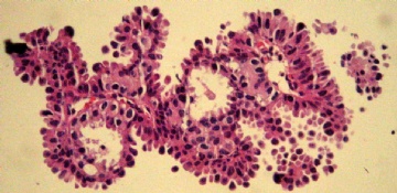
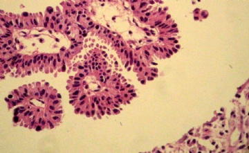
下图是以前的蜡块,重新切片染色,今天刚得到切片。

华夏病理/粉蓝医疗
为基层医院病理科提供全面解决方案,
努力让人人享有便捷准确可靠的病理诊断服务。
See H&E carefully in High power. Generally the tumor cells look a little difference compared with surrounding benign ductal cells.
In fact they can be idc based on HE, except for the glands in the left low photos (glands right side). If all myoepithelial markers are negative, they may be invasive glands. Of cause we need to consider the case individually. Are there only two or three glands without myoepithelial cells surrounding?
-
lfl001200546 离线
- 帖子:2808
- 粉蓝豆:40
- 经验:2808
- 注册时间:2007-02-14
- 加关注 | 发消息


