| 图片: | |
|---|---|
| 名称: | |
| 描述: | |
- B1820乳房不典型血管瘤( cqz-20)
| 姓 名: | ××× | 性别: | 年龄: | ||
| 标本名称: | |||||
| 简要病史: | |||||
| 肉眼检查: | |||||
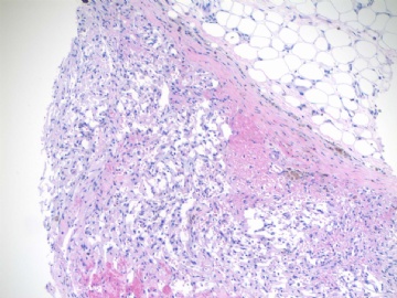
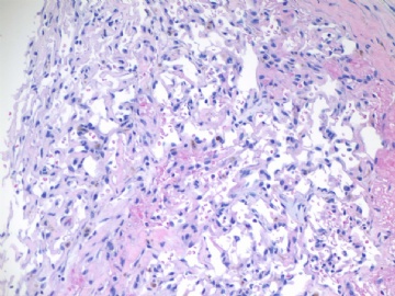
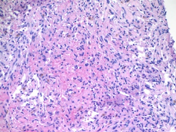
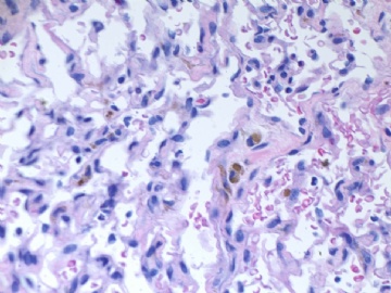
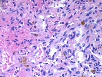
About 50 y/f breast core bx.
Immaging showed a 0.7x0.5 cm demarcated abnormal area.
F1,100x, demostrating the margins
F2-3 200x
F4-5 400x
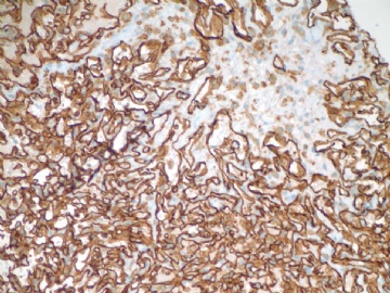
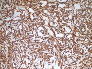
F6 CD31stain
F7 CD34 stain
-
本帖最后由 于 2009-07-18 09:31:00 编辑
相关帖子
-
本帖最后由 于 2009-07-08 22:44:00 编辑
| 以下是引用quyibl在2009-6-15 20:22:00的发言:
沉了,顶上来! KI67阳性率不超过5%,肿瘤大小0.7*0.5cm,无放射史。这些都不支持诊断恶性。没经验,搬个板凳学习中....等待老师的讲解。 |
Good. I am waiting you and others' final diagnosis.
Now you know all information for this pt. How would you sign out this case if it were your pt. Then I will tell you my report.
Of cause my report does not mean correct one.
abin译:
很好。我在等着你和别人的最终诊断。
现在你知道了患者的所有信息。如果是你的病例,你会怎样签发报告?然后我会告诉你,我是怎样签发的。
当然我的报告并不意味着一定正确。
-
似乎Ki67很有帮助,提供一些资料:
The Ki67-labeling index of angiosarcomas (mean, 38.1; median, 40.3) is substantially higher than the labeling index of hemangiomas (mean, 4.6; median, 1.7) (178). The labeling index of low-grade angiosarcomas (mean, 29.4; median, 24.5) is considerably less than the labeling indices for intermediate (mean, 41.6; median, 42.9) and high-grade angiosarcomas (mean, 44.8; median, 43.5).
The Ki67 immunostain is a useful adjunct in the diagnosis of the mammary hemangiomas. The nuclear Ki67 labelling index in mammary hemangiomas is very low, rarely exceeding 5%. Focally higher rates of labeling may be found in a hemangioma at sites of organizing thrombi or where a biopsy was previously performed on the lesion. Therefore, it is important to have an H&E-stained section available to visualize structural details when the Ki67 stain is being interpreted. The Ki67 labeling index of mammary angiosarcomas is greater than in hemangiomas, typically exceeding 20%, even in low-grade tumors. Because the distribution of labeling is not uniform in an angiosarcoma, it is possible by chance to obtain a small biopsy sample with less than 5% labelling from a low-grade angiosarcoma. A robust Ki67 labeling index on a needle core biopsy from a mammary vascular lesion would strongly favor angiosarcoma. Very sparse labelling in such a limited sample can assist in making a diagnosis of hemangioma when correlated with the H&E appearance of the lesion.
(From Rosen's Breast Pathology, 3rd Edition)
参考文献178同五楼:Shin SJ, Lesser MJ, Rosen PP. Hemangiomas and angiosarcomas of the breast: Diagnostic utility of cell cycle markers with emphasis on Ki-67. Arch Pathol Lab Med 2007;131:538–544.

华夏病理/粉蓝医疗
为基层医院病理科提供全面解决方案,
努力让人人享有便捷准确可靠的病理诊断服务。
-
本帖最后由 于 2009-07-08 22:40:00 编辑
http://www.ipathology.cn/forum/forum_display.asp?keyno=143381
Dr. wfbjwt had an interesting breast vascular lesion case above with a lot of discussion. You can compare the two cases for study purpose if you are interested.
abin译:
http://www.ipathology.cn/forum/forum_display.asp?keyno=143381
这个链接是Dr.wfbjwt提供的一例关于乳腺血管病变的有趣病例,有很多讨论。如果有兴趣,作为学习目的,你可以比较这两个病例。
-
本帖最后由 于 2009-06-09 21:45:00 编辑
Thank all discussion above. Very good. This is the way for discussion.
When we make a diagnosis we should consider the sample types. Generally our diagnosis will guide the next surgical procedure. The standard care is that the women should have total masterectomy if we call angiosarcoma.
To answer Dr. 漫游人 's question: The location of the lesion is 11 o'clock, parenchyma. You analysis is excelelnt.
Dr. Rosen's paper quyibl mentioned is a good one in term of benign or malignant breast vascular lesion.
I had ki67 stain. Will take a photo and paste here.
Welcome more people to share the oppinion. Thanks, cz
quyibl译:
谢谢上面精彩的讨论。这才是讨论的方式。
当我们做出一个诊断时我们应当考虑标本的类型。通常我们的诊断将指导下一步的外科手术方式。如果我们诊断血管肉瘤,标准的处理方式是这位女士做全乳腺切除。
回答“漫游人”医生的问题:肿块位于11点,乳腺实质内。你的分析很精彩。
Rosen的论文是一个不错的鉴别乳腺血管肿瘤良恶性的方法。我做了ki67的染色,会采集照片贴在这的。
欢迎更多的人参与讨论。谢谢,CZ。
-
本帖最后由 于 2009-07-08 22:37:00 编辑
features support benign lesion:
1. mammographically small (<1cm) and mammographically and microscopically demarcated
2. pt has no cancer or radiotherapy history
3. cytologically bland
4. no significant anastomosing channels
factors suspicious for malignancy:
1. vascular lesion in breast is always a worry
2. morphologically it does not look like any classical hemangioma
3. it has focal anastomosing channels
4. could it be the periphery of a low grade malignant angiosarcoma
features do no support a malignancy:
1. this lesion is not just small but also appears well decarcated. Angiosarcomas of both subcutaneous and breast parenchymal forms are usually larger than 2cm and are infiltrating and destructive even in low grade ones.
what to do?
1. where is the exact location of the lesion? subcutaneous or within breast parenchyma? photos did not show breast tissue.
2. i'd like to stain smooth muscle actin to see whether there is prominent pericyte surrounding channels or only incomplete or scaterred pericytes---although i'm not sure whether it's going to work. Ki67 stain--as quyibl adviced.
3. if above does not solve the problem, can i sign the report as: vascular lesion, favour benign, however, a low grade angiosarcoma can not be completely excluded. complete excision of lesion for pathology examination is recommanded.
NJWBHUANG译:
支持良性的特征:
1.乳腺摄片小(<1cm),乳腺摄片和镜下境界清楚,
2.病人无癌或放疗史
3.细胞温和
4.无明显的互相吻合的通路
怀疑恶性的因素:
1.乳腺血管病变总是比较麻烦
2.形态学上它不像任何经典的血管瘤
3.有局灶性相互沟通的血管网
4.可能史低级别恶性血管肉瘤的外周
不支持恶性的特征:
1.这个病变不仅仅小,而且也似乎是境界清楚。无论是皮肤还是乳腺实质的血管肉瘤通常大于2cm,即使是低级别也是浸润性和破坏性生长。
2.病变确切的位置在哪里?皮下还是乳腺实质内?图片未见乳腺组织。
3.我喜欢染SMA看看是否有明显的周细胞或仅有不完整或散在的周细胞——但正如quyibl建议可作Ki-67染色,尽管我不确定是否有帮助。
4.如果做了上面的工作仍然不能做出明确诊断,我会发血管性病变,倾向良性。然后,低级别血管肉瘤不能完全排除。建议完整切除送检。
1: Am J Surg Pathol. 1991 Dec;15(12):1130-5
Sinusoidal hemangioma. A distinctive benign vascular neoplasm within the group of cavernous hemangiomas.
Department of Histopathology, St. Thomas's Hospital, London, England.
Twelve cases of sinusoidal hemangioma, a distinctive subset of the group of lesions known as cavernous hemangioma, are described. All presented as solitary subcutaneous/deep dermal lesions in adults, predominantly females. Five arose on a limb and five on the trunk; two of the latter were situated in mammary subcutaneous tissue. Histologically they were characterized by dilated, interconnecting, thin-walled vascular channels that frequently showed a pseudopapillary pattern. These vessels had a predominantly lobular architecture but peripherally showed focally ill-defined spread into subcutaneous tissue. The lining endothelium was single-layered but showed focal pleomorphism and hyperchromasia, which, combined with the pseudopapillae and apparent infiltrative pattern in areas, raised the possibility of angiosarcoma in four cases, most notably in the breast lesions. This possibility was further suggested by the presence of pseudonecrotic central infarction in two cases. Follow-up in eight cases, however, has revealed no tendency for either local recurrence or metastasis.
PMID: 1746680 [PubMed - indexed for MEDLINE]
不知道是否符合这个。
-
本帖最后由 于 2009-06-07 08:50:00 编辑
利用网络学习:
本例为血管源性肿瘤应该没什么疑问了,关键是报血管瘤还是血管肉瘤,组织学上血管瘤和Ⅰ级血管肉瘤是有重叠的,本例没有放射史和乳腺癌病史,就更增加了诊断的难度。搜素网络上的相关研究,发现一研究用ki-67来辅助区别:
Hemangiomas and angiosarcomas of the breast: diagnostic utility of cell cycle markers with emphasis on Ki-67.(Clinical report)细胞周期标记物尤其是ki-67在诊断乳腺血管瘤和血管肉瘤中的作用。
链接:http://www.accessmylibrary.com/coms2/summary_0286-32184721_ITM
|
Hemangiomas and angiosarcomas of the breast: diagnostic utility of cell cycle markers with emphasis on Ki-67.(Clinical report)
Publication: Archives of Pathology & Laboratory Medicine Publication Date: 01-APR-07 Author: Shin, Sandra J. ; Lesser, Martin ; Rosen, Paul Peter Context.--Vascular tumors comprise a minor subgroup of tumors arising in the breast and represent variants of hemangiomas and angiosarcomas. Diagnostic challenges may arise when differentiating hemangiomas from types I and II angiosarcomas. Ki-67 expression has been used as an adjunct to distinguish between benign and malignant lesions exhibiting histologic overlap at various anatomic sites. angiosarcomas, positivity for Ki-67 was inversely related to that of p27 but not to Skp2 or cyclin D1. This was also true among hemangiomas. | ||

- 我思故我在! know something about everything,know everything about something.
-
本帖最后由 于 2009-06-07 17:47:00 编辑
I remeber we had a breast vascular lesion case in this web with a lot of debatable discusion.
Above is a my case three weeks ago. Patient has no cancer or radiation history. She did have a breast core biopsy in the same breast two years ago. The diagosis was small papilloma and ducatal hyperplsia. I reviewed the previous breast core slides and agree with the interpretation. I called the radiologist to review the current and previous imaging films to confirm current lesion and the biopsy lesion two years ago were different areas. I felt difficult to sign out the breasst core biopsy case. Just wonder how you guys would sign out the case if it were yours.
I am on service for FNA on site evaluation for surgeons (several lung mass patients) this weekend. Now we are waiting for patient and I have time to send you this case. Enjoy your weekend
cz
quyibl译:
我记得网上有过一个存在很大争议的乳腺血管性病变的病例。
上面这个是我三周前的一个病例。病人没有乳腺癌或放射线照射史。两年前同侧乳腺做过活检,诊断是小的乳头状瘤伴导管增生。我复习了原先的乳腺活检切片,同意当时的诊断。我请放射科医师复验现在和原先的影像片,确定两次病变不在同一位置。我感到签出这个病例很困难,非常想知道如果这是你的病例,你会怎么签发这个报告。
这个周末, 我在为外科医生做现场的FNA(几个肺部包块的病人),现在正在等待病人,所以有时间发送给你们这个病例。 周末愉快!cz
