| 图片: | |
|---|---|
| 名称: | |
| 描述: | |
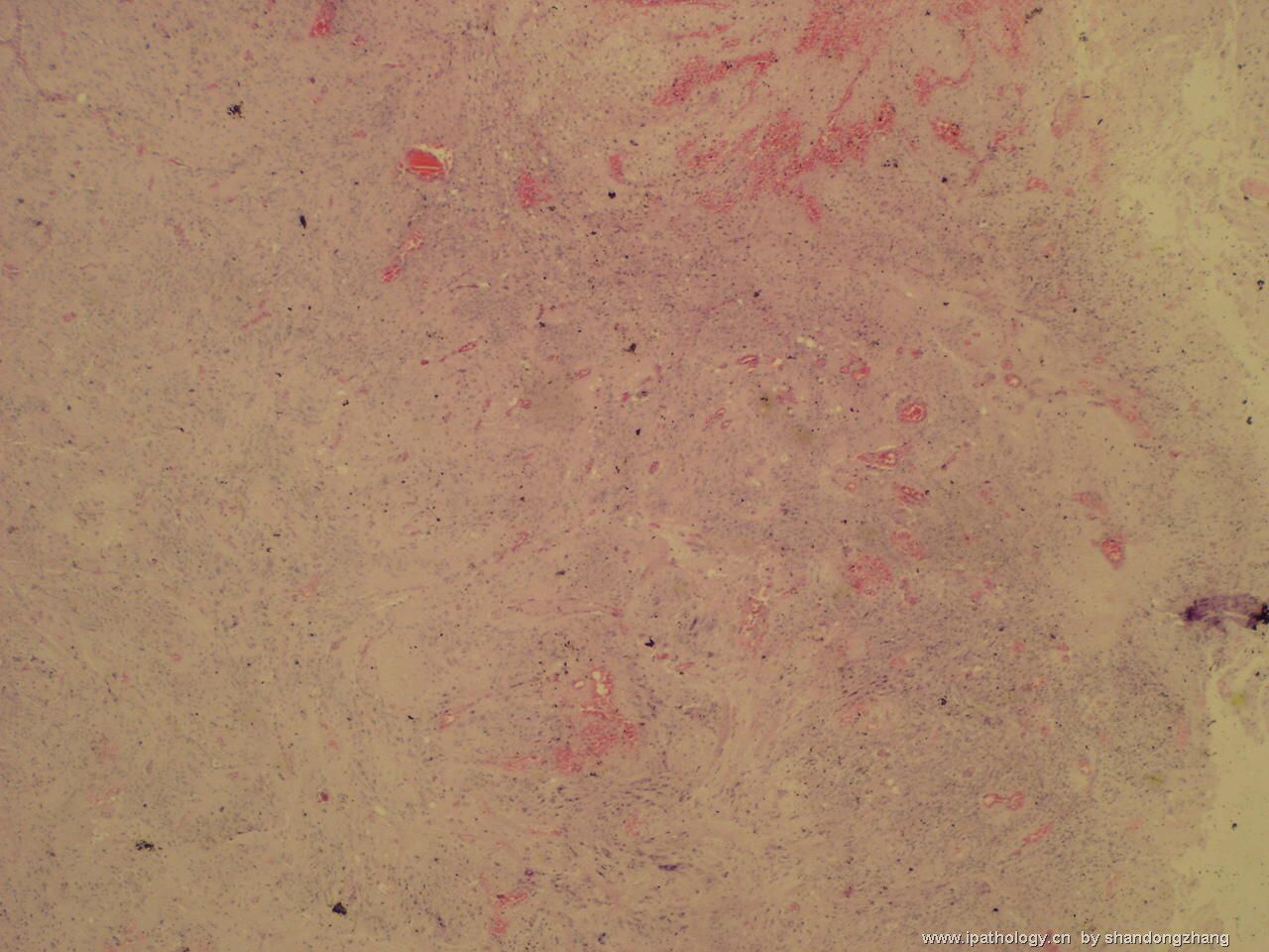
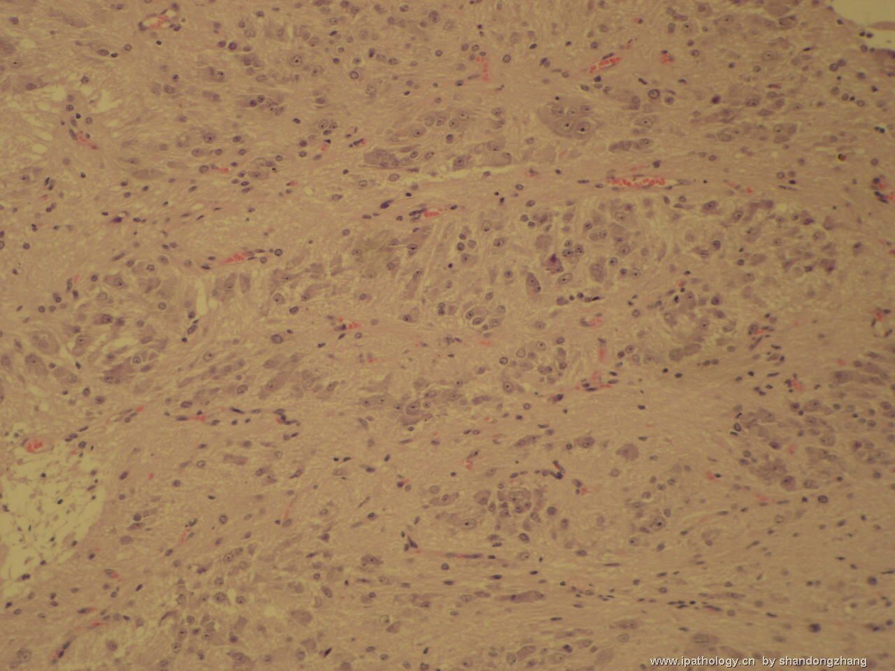
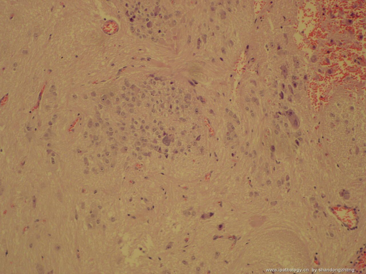
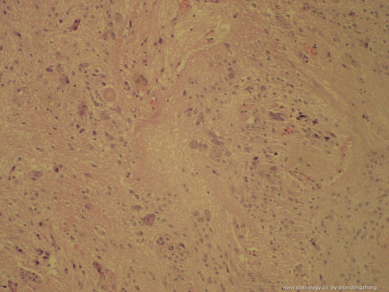
- 左颞叶肿瘤
-
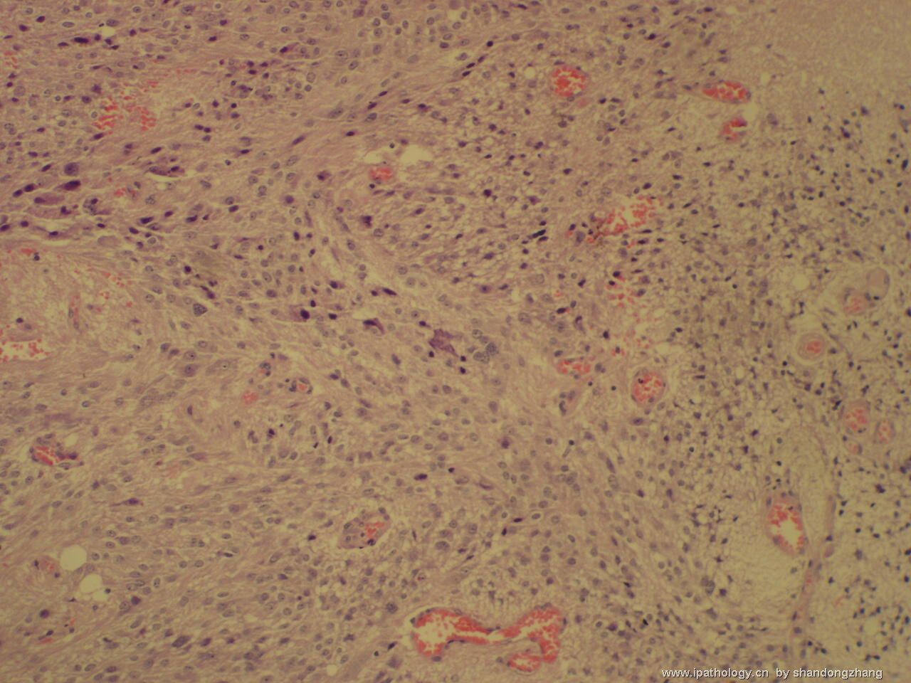
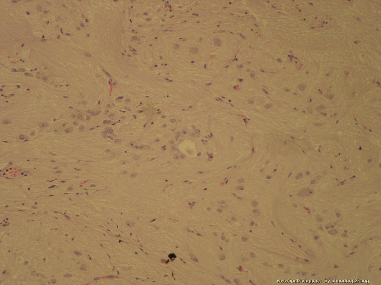
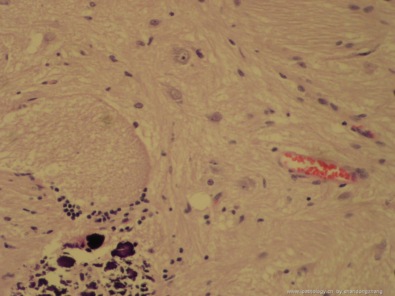
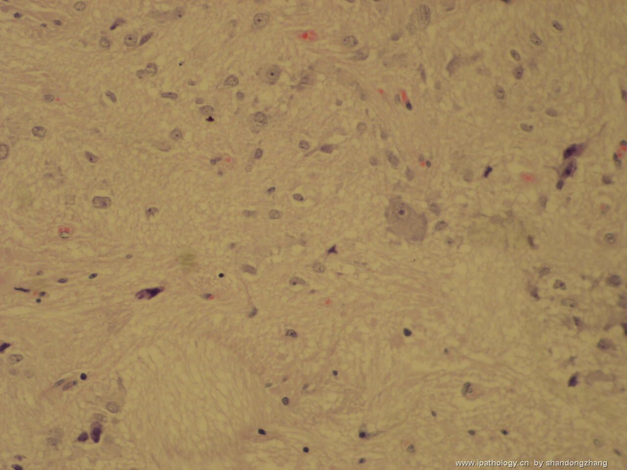
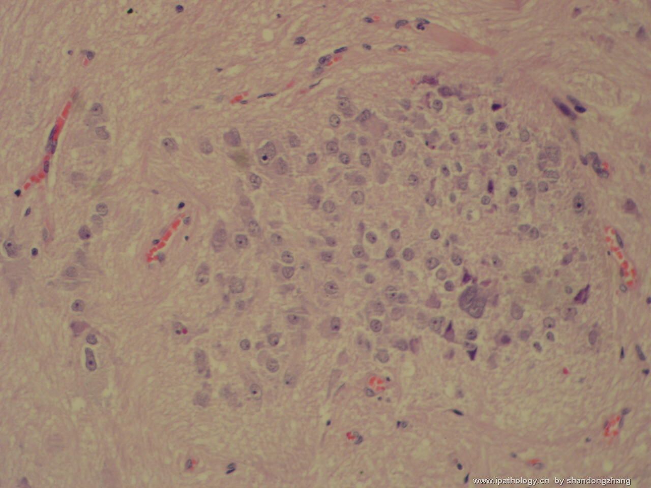
Though Figure No 5 is cellular, most cells in this neoplasm have cytologic features of ganglion cells or neurons. Calcification is seen as well. That make this a WHO grade I gangliocytoma. I do not see a gliaoma component to make this a ganglioglioma. In general, WHO grade I gangliocytomas are benign lesions found mainly during workup of seizure disorders. Grading of a ganglioglioma depends on the co-existing glioma (usually astrocytoma) present. It could be grade I (if the glioma is a pilocytic astrocytoma), grade II (diffuse or fibrillary astrocytoma), grade III (anaplastic astrocytoma) or, very rarely, grade IV (glioblastoma). Sometimes, differentiated ganglion cells are found with primitive embryonal cells that also express neuronal markers (NeuN, NSE. synaptophysin, neurofilaments). That combination qualifies the lesion as a WHO grade IV ganglioneuroblastoma.

聞道有先後,術業有專攻
First and importantly, this case is not 发育不良性小脑节细胞瘤(WHO grade I dysplastic cerebellar gangliocytoma or Lhermitte-Duclos disease) that has very specific gross and histopathology, and is sometimes associated with Cowden disease. Figure 1 shows possible necrosis in the congested corner, but this is not associated with features of a high grade neoplasm. Grade III anaplastic ganglioglioma, grade IV glioblastoma arising from a ganglioglioma, and grade IV ganglioneuroblastoma are not difficult to diagnose.
Distinction between grade I gangliocytoma and grade I ganglioglioma is probably not an important one, since they have similar if not identical biologic behavior and prognosis. Certainly, one has to see a gliomatous component to call a neuronal tumor a ganglioglioma. This judgment can be very subjective - "all in the eyes of the beholder". From the photos you kindly provided, I have not seen a definite gliomatous component in this case. Short of seeing relatively large areas (greater than one 100x microscopic field) of glial neoplasm without admixed dysplastic neurons, I usually would not call it a ganglioglioma. This is my personal criterion and by no means scientific. Naturally, I cannot be absolutely certain about this point without examining the histologic slides myself.
The distinction between WHO grade I and grade II gangliogliomas is not as clear cut as it seems. If one uses the conventional definitions, grade I ganglioglioma is very uncommon. Remember, pure gangliocytomas often contain eosinophilic granular bodies. Therefore, one would like to see other characteristic features of pilocytic astrocytoma (biphasic growth pattern, mucinous microcysts, Rosenthal fibers, uniform piloid cells) before calling any tumor grade I ganglioglioma. In the photos uploaded so far, I have not seen these features. Finally, when the gliomatous component is very bland and I cannot decide, I would just diagnose it as a grade I~II ganglioglioma. I hope my comment is more enlightening than confusing.

聞道有先後,術業有專攻
-
Though Figure No 5 is cellular, most cells in this neoplasm have cytologic features of ganglion cells or neurons. Calcification is seen as well. That make this a WHO grade I gangliocytoma. I do not see a gliaoma component to make this a ganglioglioma. In general, WHO grade I gangliocytomas are benign lesions found mainly during workup of seizure disorders. Grading of a ganglioglioma depends on the co-existing glioma (usually astrocytoma) present. It could be grade I (if the glioma is a pilocytic astrocytoma), grade II (diffuse or fibrillary astrocytoma), grade III (anaplastic astrocytoma) or, very rarely, grade IV (glioblastoma). Sometimes, differentiated ganglion cells are found with primitive embryonal cells that also express neuronal markers (NeuN, NSE. synaptophysin, neurofilaments). That combination qualifies the lesion as a WHO grade IV ganglioneuroblastoma.
虽然图5为细胞的,此肿物中的大多数细胞具有神经节细胞或神经元的细胞特点,钙化也可见到。根据以上特点,可诊断此肿物为一个WHOⅠ级神经节细胞瘤。我没有看到胶质瘤的成分,故不能诊断节细胞胶质瘤。一般来说,WHOⅠ级神经节细胞瘤是良性病变,主要在癫痫发作做病情检查时发现。节细胞胶质瘤的分级主要依赖于并存的神经胶质瘤(通常是星形细胞瘤)。Ⅰ级(神经胶质瘤为毛细胞星形细胞瘤),Ⅱ级(弥散的或纤维星形细胞瘤),Ⅲ级(间变型星形细胞瘤),Ⅳ级非常少见(胶质母细胞瘤)。有时,分化好的神经节细胞与也可表达神经标记(NeuN,NSE,syn,NF)的原始胚胎性细胞并存。这两种细胞合并存在的现象足以诊断此病变为WHOIV 级成神经节胶质细胞瘤。
-
At the request of the author Dr. Zhang, I read the discussion on the same case at CPN. Thank you for allowing me to address this case further.
First and importantly, this case is not 发育不良性小脑节细胞瘤(WHO grade I dysplastic cerebellar gangliocytoma or Lhermitte-Duclos disease) that has very specific gross and histopathology, and is sometimes associated with Cowden disease. Figure 1 shows possible necrosis in the congested corner, but this is not associated with features of a high grade neoplasm. Grade III anaplastic ganglioglioma, grade IV glioblastoma arising from a ganglioglioma, and grade IV ganglioneuroblastoma are not difficult to diagnose.
应作者张大夫之要求,我看了中华病理学网上关于此病例的讨论。谢谢你让我对此病例作进一步分析。首先最重要的是,此病例不是发育不良性小脑节细胞瘤(WHOⅠ级小脑发育不良性节细胞瘤又名Lhermitte-Duclos病),发育不良性小脑节细胞瘤具有非常特殊的大体和组织学表现,有时与考登病相关。图1显示充血区域可能有一些坏死,但这并不是高分化肿瘤的特点。Ⅲ级未分化的节细胞胶质瘤、Ⅳ级胶质母细胞瘤起因于节细胞胶质瘤,Ⅳ级成神经节胶质细胞瘤也不难诊断。
-
Distinction between grade I gangliocytoma and grade I ganglioglioma is probably not an important one, since they have similar if not identical biologic behavior and prognosis. Certainly, one has to see a gliomatous component to call a neuronal tumor a ganglioglioma. This judgment can be very subjective - "all in the eyes of the beholder". From the photos you kindly provided, I have not seen a definite gliomatous component in this case. Short of seeing relatively large areas (greater than one 100x microscopic field) of glial neoplasm without admixed dysplastic neurons, I usually would not call it a ganglioglioma. This is my personal criterion and by no means scientific. Naturally, I cannot be absolutely certain about this point without examining the histologic slides myself.
区分Ⅰ级神经节细胞瘤和Ⅰ级节细胞胶质瘤可能不是一个重要问题,因为它们的生物学行为和预后即便不完全相同也很相似。当然,必须看到胶质瘤成分才能诊断一个神经肿瘤为节细胞胶质瘤,这一判断非常主观--"亡斧疑邻"。从你热心提供的照片中,我并没有看到明确的胶质瘤成分。缺乏相对大区域(大于100倍显微镜视野)不混合存在分化不良神经元成分的胶质瘤成分,我通常不诊断为节细胞胶质瘤。这个是我个人的诊断标准并不具有科学性。没有亲自观察切片,我对此观点也没有绝对把握。
-
The distinction between WHO grade I and grade II gangliogliomas is not as clear cut as it seems. If one uses the conventional definitions, grade I ganglioglioma is very uncommon. Remember, pure gangliocytomas often contain eosinophilic granular bodies. Therefore, one would like to see other characteristic features of pilocytic astrocytoma (biphasic growth pattern, mucinous microcysts, Rosenthal fibers, uniform piloid cells) before calling any tumor grade I ganglioglioma. In the photos uploaded so far, I have not seen these features. Finally, when the gliomatous component is very bland and I cannot decide, I would just diagnose it as a grade I~II ganglioglioma. I hope my comment is more enlightening than confusing.
WHOⅠ级和Ⅱ级节细胞胶质瘤的差别并不象划分的那么明确。如果按照传统的限定标准,Ⅰ级节细胞胶质瘤不常见。需要记住的一点是,单纯的神经节细胞瘤通常含有嗜酸性颗粒小体。因此,人们在诊断任何一个肿瘤为Ⅰ级节细胞胶质瘤之前,通常看一下它是否具备纤维星形细胞瘤的其它特点(双相生长方式,粘蛋白样微囊,Rosenthal纤维,均匀一致的纤维性细胞)。到目前为止上传的图片中,我没有看到这些特点。最后,如果这些胶质瘤成分非常温和而我不能判定时,我一般诊断为Ⅰ-Ⅱ级节细胞胶质瘤。我希望我的上述陈述能有一些启发作用,而不会让人更加困惑。
| 以下是引用lff 在2006-11-1 17:03:00的发言: Though Figure No 5 is cellular, most cells in this neoplasm have cytologic features of ganglion cells or neurons. Calcification is seen as well. That make this a WHO grade I gangliocytoma. I do not see a gliaoma component to make this a ganglioglioma. In general, WHO grade I gangliocytomas are benign lesions found mainly during workup of seizure disorders. Grading of a ganglioglioma depends on the co-existing glioma (usually astrocytoma) present. It could be grade I (if the glioma is a pilocytic astrocytoma), grade II (diffuse or fibrillary astrocytoma), grade III (anaplastic astrocytoma) or, very rarely, grade IV (glioblastoma). Sometimes, differentiated ganglion cells are found with primitive embryonal cells that also express neuronal markers (NeuN, NSE. synaptophysin, neurofilaments). That combination qualifies the lesion as a WHO grade IV ganglioneuroblastoma. |

聞道有先後,術業有專攻
| 以下是引用lff 在2006-11-1 17:09:00的发言: At the request of the author Dr. Zhang, I read the discussion on the same case at CPN. Thank you for allowing me to address this case further. |

聞道有先後,術業有專攻
| 以下是引用lff 在2006-11-1 17:14:00的发言: Distinction between grade I gangliocytoma and grade I ganglioglioma is probably not an important one, since they have similar if not identical biologic behavior and prognosis. Certainly, one has to see a gliomatous component to call a neuronal tumor a ganglioglioma. This judgment can be very subjective - "all in the eyes of the beholder". From the photos you kindly provided, I have not seen a definite gliomatous component in this case. Short of seeing relatively large areas (greater than one 100x microscopic field) of glial neoplasm without admixed dysplastic neurons, I usually would not call it a ganglioglioma. This is my personal criterion and by no means scientific. Naturally, I cannot be absolutely certain about this point without examining the histologic slides myself. |

聞道有先後,術業有專攻
| 以下是引用lff 在2006-11-1 17:19:00的发言: The distinction between WHO grade I and grade II gangliogliomas is not as clear cut as it seems. If one uses the conventional definitions, grade I ganglioglioma is very uncommon. Remember, pure gangliocytomas often contain eosinophilic granular bodies. Therefore, one would like to see other characteristic features of pilocytic astrocytoma (biphasic growth pattern, mucinous microcysts, Rosenthal fibers, uniform piloid cells) before calling any tumor grade I ganglioglioma. In the photos uploaded so far, I have not seen these features. Finally, when the gliomatous component is very bland and I cannot decide, I would just diagnose it as a grade I~II ganglioglioma. I hope my comment is more enlightening than confusing. |

聞道有先後,術業有專攻
-
本帖最后由 于 2006-11-11 12:12:00 编辑
应部分朋友的要求,将lff翻译整理如下:
第2楼,mjma老师的点评:
尽管图5细胞丰富,此肿瘤中的大多数细胞仍具有神经节细胞或神经元的细胞特点,钙化也可见到。根据以上特点,可诊断此肿物为一个WHOⅠ级神经节细胞瘤。我没有看到胶质瘤的成分,故不能诊断节细胞胶质瘤。一般来说,WHOⅠ级神经节细胞瘤是良性病变,主要在癫痫发作查见。节细胞胶质瘤的分级主要依赖于并存的胶质瘤(通常是星形细胞瘤)。Ⅰ级(胶质瘤为毛细胞型星形细胞瘤),Ⅱ级(弥散性或纤维型星形细胞瘤),Ⅲ级(间变型星形细胞瘤),或者非常少见的Ⅳ级(胶质母细胞瘤)。有时,分化好的神经节细胞与也可表达神经标记(NeuN,NSE,syn,NF)的原始胚胎性细胞并存。这两种细胞合并存在的现象足以诊断此病变为WHOIV 级神经节神经母细胞瘤。
第4楼,mjma老师的点评:
应Dr. Zhang要求,我看了中国病理学网上关于此病例的讨论。谢谢你让我对此病例作进一步分析。首先最重要的是,此病例不是发育不良性小脑节细胞瘤(WHOⅠ级小脑发育不良的节细胞瘤又名Lhermitte-Duclos病),此瘤具有非常特殊的大体和组织学表现,有时与Cowden综合征(译注:伴有多发性错构瘤-肿瘤综合征)相关。图1显示充血的一角可能有坏死,但这与高级别肿瘤的特点无关。Ⅲ级间变性节细胞胶质瘤、起源于节细胞胶质瘤的Ⅳ级胶质母细胞瘤,以及Ⅳ级神经节神经母细胞瘤也不难诊断。
区分Ⅰ级神经节细胞瘤和Ⅰ级节细胞胶质瘤可能并不重要,因为它们的生物学行为和预后即便不完全相同也很相似。当然,必须看到胶质瘤成分才能将神经肿瘤诊断为节细胞胶质瘤。这一判断非常主观--"亡斧疑邻"。从你热心提供的照片中,我并没有看到此例有明确的胶质瘤成分。缺乏相当大区域(大于100倍显微镜视野)的胶质瘤成分,不伴混合性发育不良的神经元成分,我通常不会称之为节细胞胶质瘤。这个是我的个人标准,并不科学。当然,在没有亲自观察切片的前提下,我也不能完全确诊。
WHOⅠ级和Ⅱ级节细胞胶质瘤的区别并不那么明确。如果按照传统的定义,Ⅰ级节细胞胶质瘤非常少见。请记住,纯粹的神经节细胞瘤通常也含有嗜酸性颗粒小体。因此,在诊断任何肿瘤为Ⅰ级节细胞胶质瘤之前,会观察毛细胞型星形细胞瘤的其它特点(双相生长,粘液性微囊,Rosenthal纤维,一致的毛细胞)。到目前为止上传的图片中,我没有看到这些特点。最后,如果这些胶质瘤成分很不明显、无法判断,我只好诊断为Ⅰ-Ⅱ级节细胞胶质瘤。我希望这些点评能有一些启发作用,而不会让人更加困惑。
第6楼,xiaohl老师的点评:
倾向于节细胞胶质瘤

华夏病理/粉蓝医疗
为基层医院病理科提供全面解决方案,
努力让人人享有便捷准确可靠的病理诊断服务。

