| 图片: | |
|---|---|
| 名称: | |
| 描述: | |
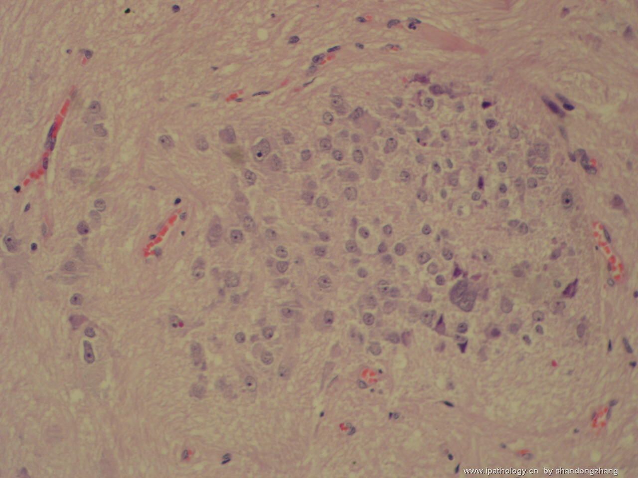
- 左颞叶肿瘤
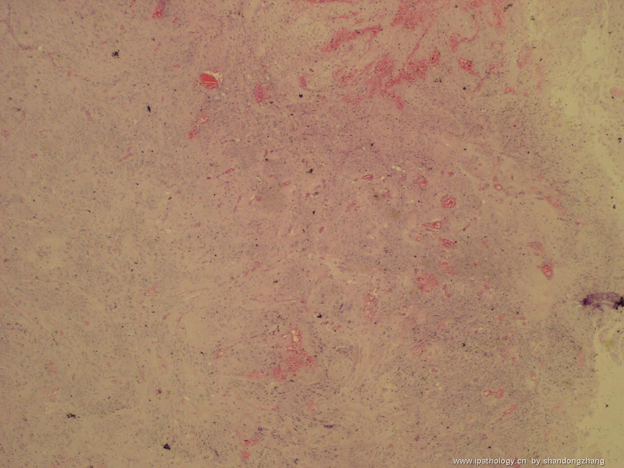
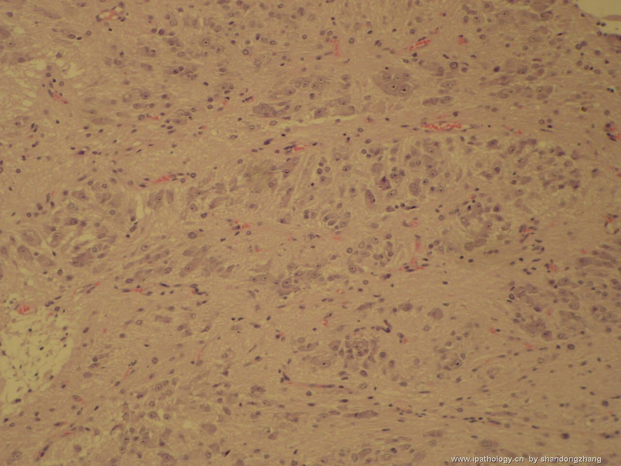
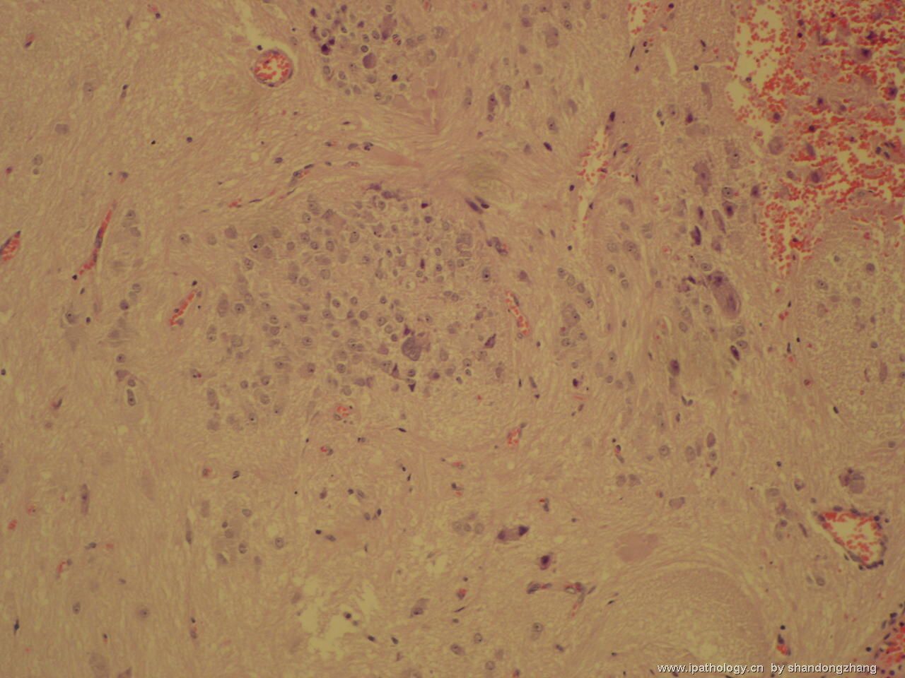
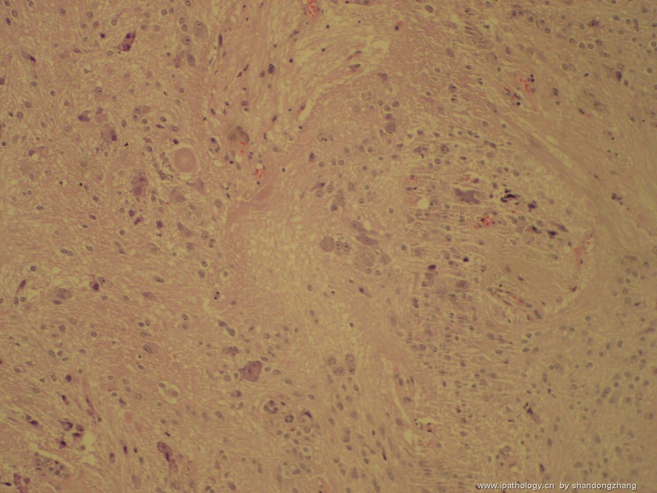
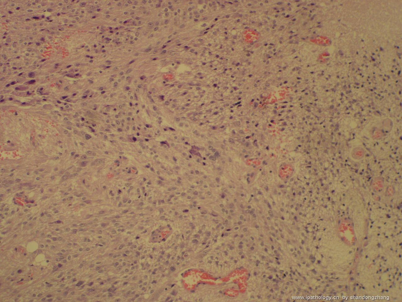
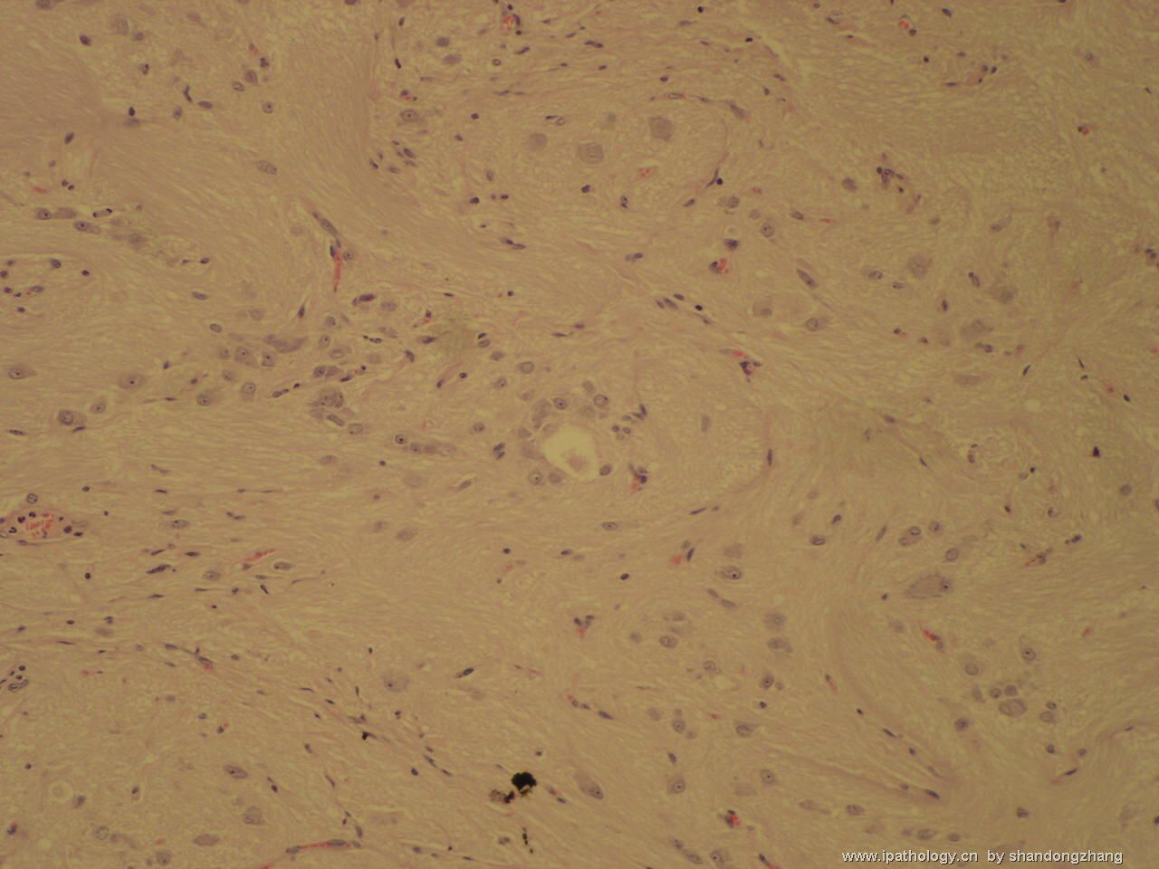
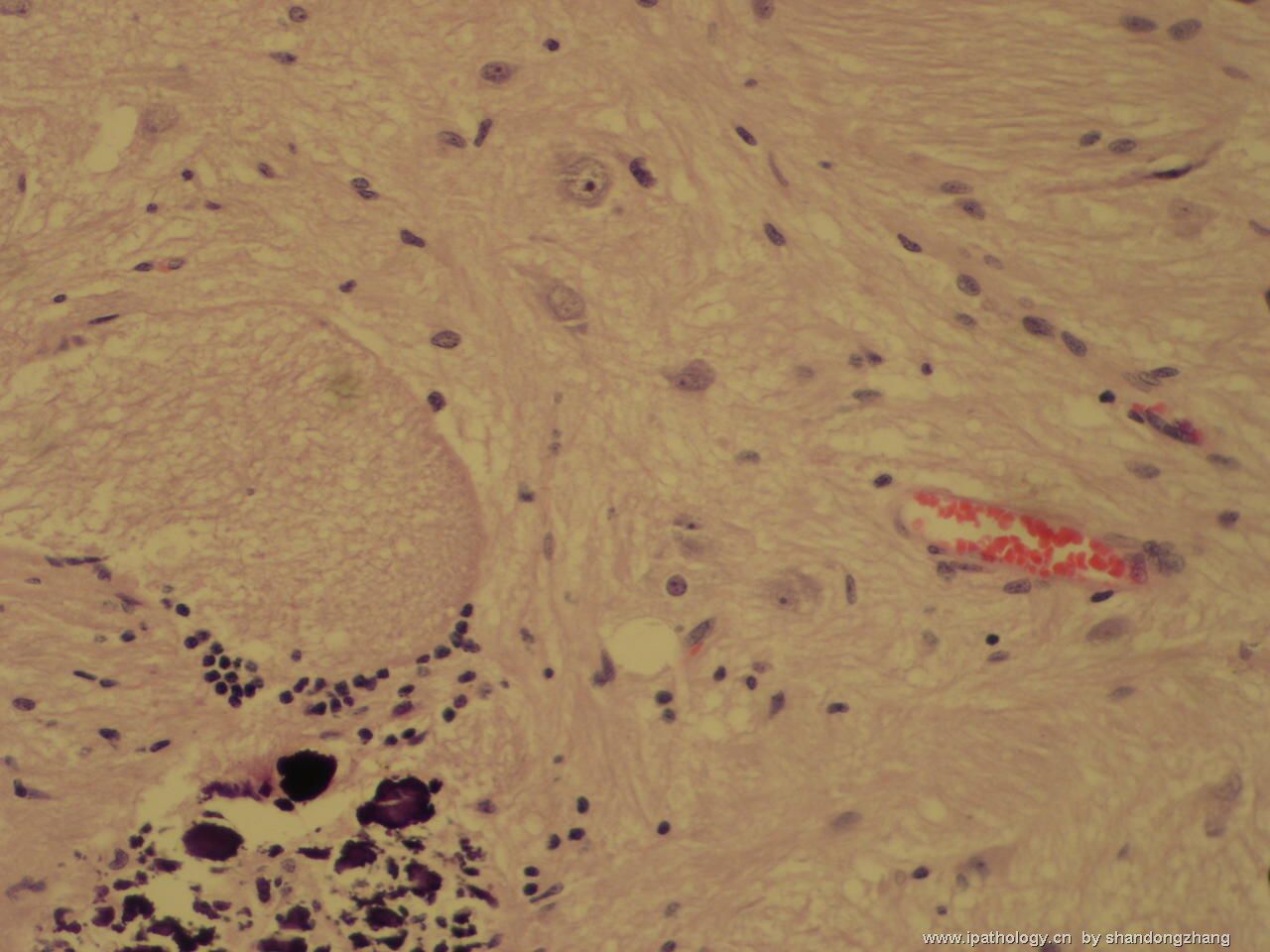
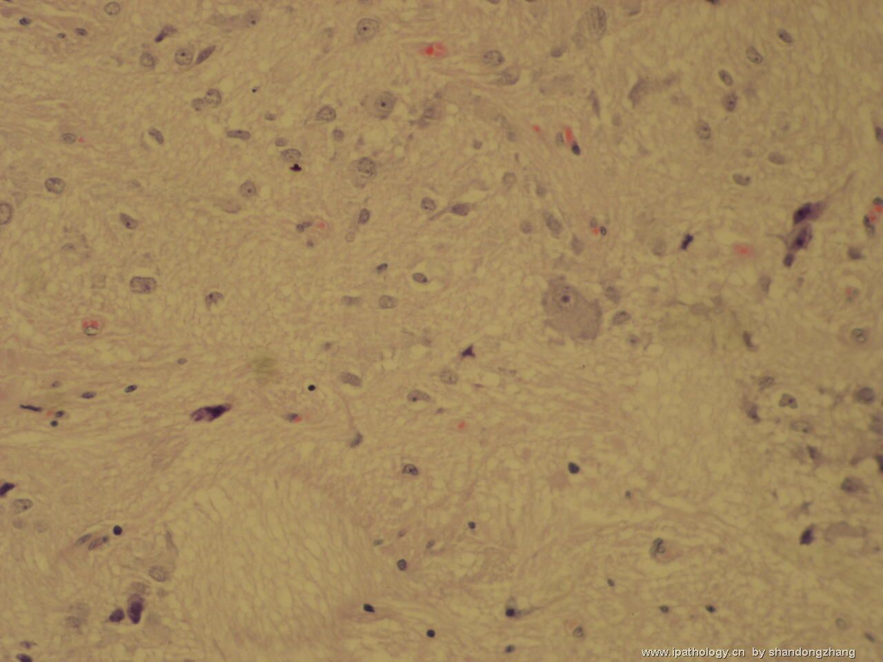
| 以下是引用mjma 在2006-10-7 13:24:00的发言: Though Figure No 5 is cellular, most cells in this neoplasm have cytologic features of ganglion cells or neurons. Calcification is seen as well. That make this a WHO grade I gangliocytoma. I do not see a gliaoma component to make this a ganglioglioma. In general, WHO grade I gangliocytomas are benign lesions found mainly during workup of seizure disorders. Grading of a ganglioglioma depends on the co-existing glioma (usually astrocytoma) present. It could be grade I (if the glioma is a pilocytic astrocytoma), grade II (diffuse or fibrillary astrocytoma), grade III (anaplastic astrocytoma) or, very rarely, grade IV (glioblastoma). Sometimes, differentiated ganglion cells are found with primitive embryonal cells that also express neuronal markers (NeuN, NSE. synaptophysin, neurofilaments). That combination qualifies the lesion as a WHO grade IV ganglioneuroblastoma. |

