| 图片: | |
|---|---|
| 名称: | |
| 描述: | |
- B1739职工家属的宫内膜病变难住了我
女 75岁
病史:绝经30年,阴道少量出血3天。乳腺癌术后2年余,口服他莫昔芬1年,现已停药1年余。
手术所见:宫腔深9厘米,宫内不平感,较硬。
彩超:宫腔内多个小结节,大的6*5毫米,无回声。
巨检:灰褐色碎组织一堆,共直径0.8厘米。
-
本帖最后由 于 2009-05-26 21:42:00 编辑
相关帖子
-
本帖最后由 于 2009-05-27 21:27:00 编辑
|
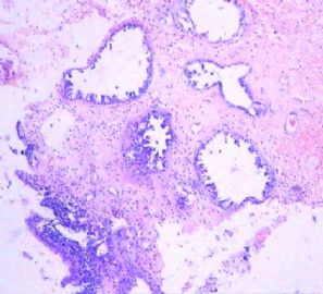
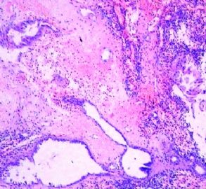
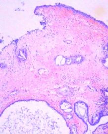
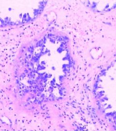
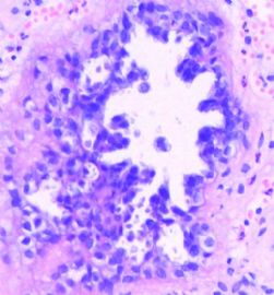
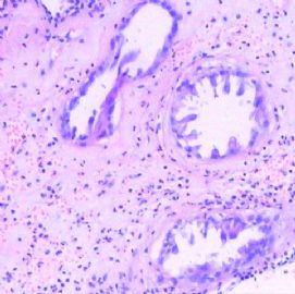
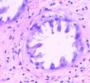
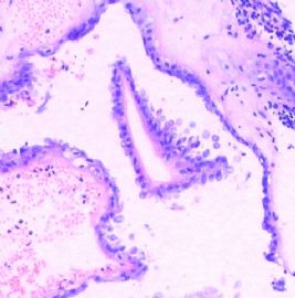
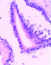
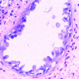
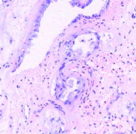
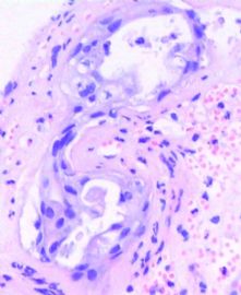
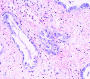
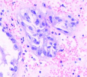
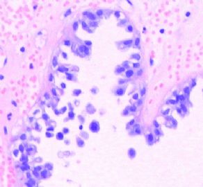
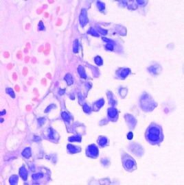
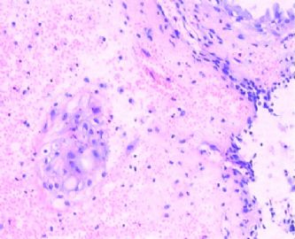
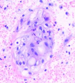
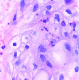
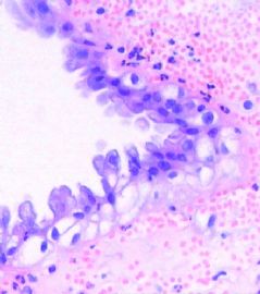
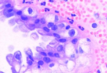
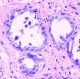
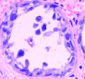
Thinking the case and check the phtos again.再次思考本例并查看图像,Agree with Dr. Yu. 同意YU大夫的意见,1. Endometrial polyp.1、子宫内膜息肉,2. Habnail pattern, may be habnail metaplasia.2、鞋钉样结构,可能是鞋钉样细胞化生,3. The clear cell change may be Arias-Stella-like change related to the tamoxifen treatment. 3、透明细胞改变可能是与他莫昔芬治疗有关的Arias-Stella样改变。Arias-Stella-like change can be mimic of adenocarcinoma, especially clear cell carcinoma. Arias-Stella样改变可以类似于腺癌,特别是类似与透明细胞癌,So I will choose endometrial polyp with Arias-Stella change if it is a question.所以,如果是考试题,我选择子宫内膜息肉伴有Arias-Stella改变。But in the true case I may still call polyp with atypical gland proliferation. 但是如果是真实病例,我仍会称为息肉伴有非典型腺体增生。Then write the comment with differential dx, and favoring dx然后再写上备注,包括鉴别诊断和倾向性诊断。Anyway, p53 and ki67 may be helpful.无论如何p53 和 ki67是可能有帮助的。 赵大夫 |
Difficult case.
Basically I agree with Abin's oppinion. The morphology does not like typical metastatic breast ca. You can review the breast ca case and compare the morphologic features.
H@E shows focal glandular proliferation with hobnail pattern and clear cytoplasm. Is this serous carcinoma or serous endometrial intraepithelial ca? It does not look like. Do IHC for p53 and WT1. Serous tumor should be strongly and diffusely positive for them, especially for P53. Of cause Clear cell ca can be positive also.
My feeling is that the lesion is a clear cell lesion. Clearly they are clear cells, hobnail growths. Nuclei are large in high power even though they are not so urgly.
Now the queation is that it is reactive change to hormon or neoplastic lesion. I have difficulty to make the judgment based on the photos. It is true it is not a classic picture of clear cell carcinoma, but i cannot completely exclude.
You can call atypical glandular proliferation with clear cell feature and suggest to get more tissue for definite diagnosis.
Just suggestion.
cz
| 以下是引用zzz333858在2009-5-27 20:30:00的发言:
5月份在北京开妇产临床病理诊断学习班,听郑文军老师讲EIC(子宫内膜癌前病变),分两型,Ⅰ型与激素有关,Ⅱ型与P53突变有关,经常是息肉样病变组织内见异型增生的腺体,P53阳性,我觉得这个可能也是吧,如果没进一步处理可能就会发生浆液性癌。 有兴趣可搜索WENJIN ZHENG PATHOLOGY,他在个人空间有讲课的材料 |
补充一些:
子宫内膜的癌前病变包括EIN和EIC。EIN为Ⅰ型癌的前驱病变,与雌激素等因素有关。EIC为Ⅱ型癌(主要是浆液性癌)的前驱病变,与P53突变有关。

华夏病理/粉蓝医疗
为基层医院病理科提供全面解决方案,
努力让人人享有便捷准确可靠的病理诊断服务。
学习了各位的观点,同意楼主的诊断。
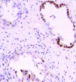
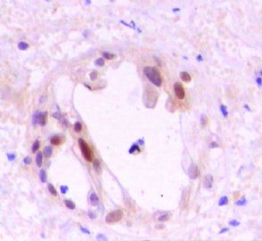
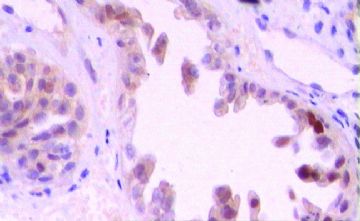
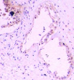
特别注意到楼主提供的免疫组化结果,Ki67灶性高增殖指数,P53也有少数细胞阳性,提示存在异型增生(郑文新教授称为“子宫内膜腺体异型增生endometrial glandular dysplasia, EmGD”),不是单纯的化生性改变。
郑文新教授认为,EmGD是介于萎缩性静止期子宫内膜与EIC之间的改变,EmGD是真正意义上的癌前病变,而EIC属于早期癌。EmGD的临床处理资料有限。本例有高危因素(乳腺癌史),如果能耐受手术,建议切除子宫。

华夏病理/粉蓝医疗
为基层医院病理科提供全面解决方案,
努力让人人享有便捷准确可靠的病理诊断服务。
-
xianyuanqq82 离线
- 帖子:455
- 粉蓝豆:55
- 经验:456
- 注册时间:2009-02-04
- 加关注 | 发消息

