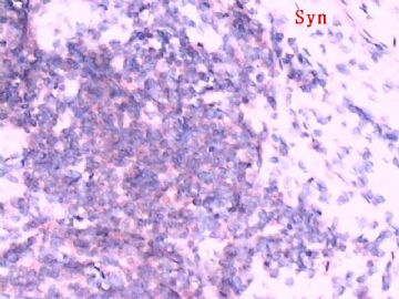
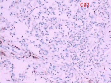
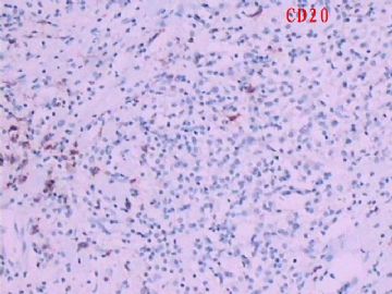
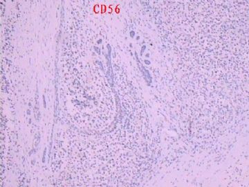
| 图片: | |
|---|---|
| 名称: | |
| 描述: | |
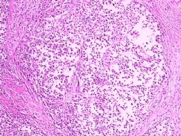
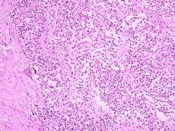
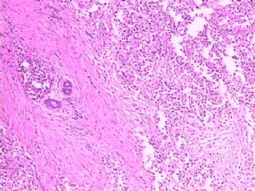
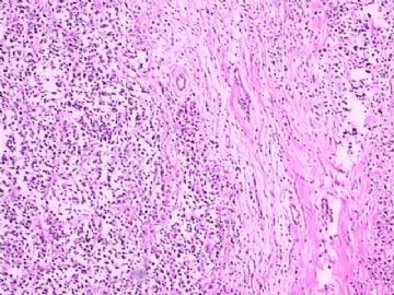
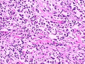
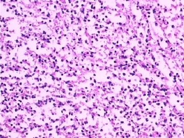
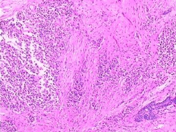
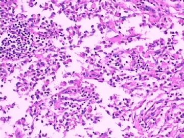
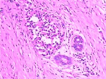
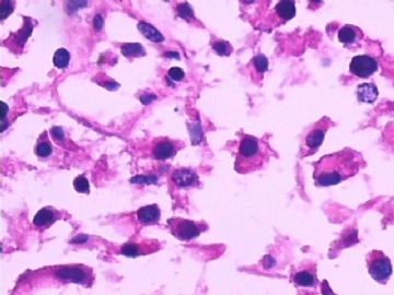
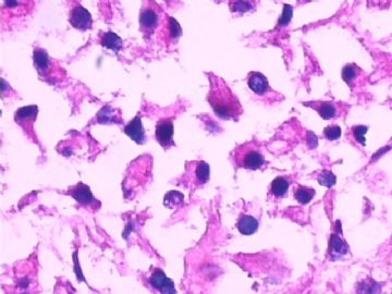
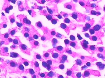
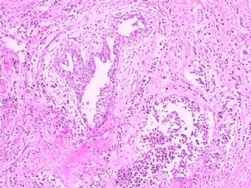
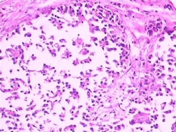
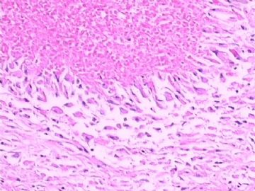
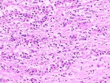
- B1783左乳肿物(罕见病例)
| 姓 名: | ××× | 性别: | 女 | 年龄: | 50 |
| 标本名称: | 左乳腺肿物 | ||||
| 简要病史: | |||||
| 肉眼检查: | 乳腺肿物,直径2cm | ||||
标签:乳腺浸润性导管癌 标本固定

- 把我们应该做的做到最好!
相关帖子
×参考诊断
-
liguoxia71 离线
- 帖子:4174
- 粉蓝豆:3122
- 经验:4677
- 注册时间:2007-04-01
- 加关注 | 发消息
-
liguoxia71 离线
- 帖子:4174
- 粉蓝豆:3122
- 经验:4677
- 注册时间:2007-04-01
- 加关注 | 发消息
-
本帖最后由 于 2009-04-24 23:14:00 编辑
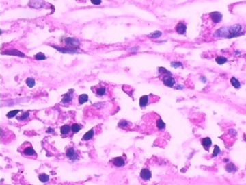
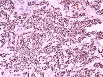
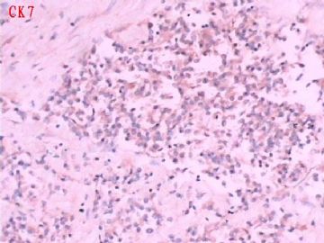
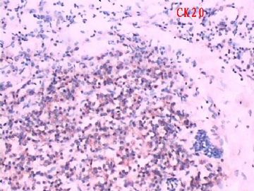
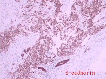
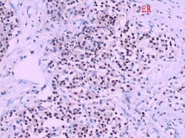
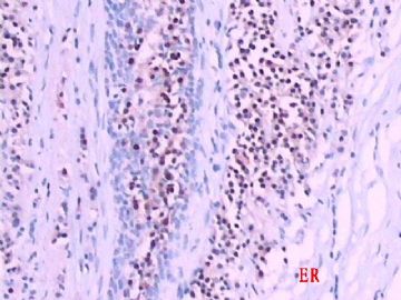
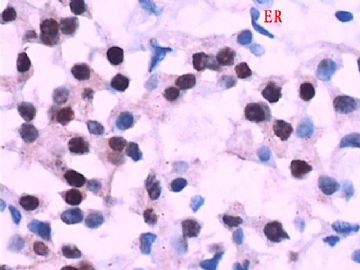
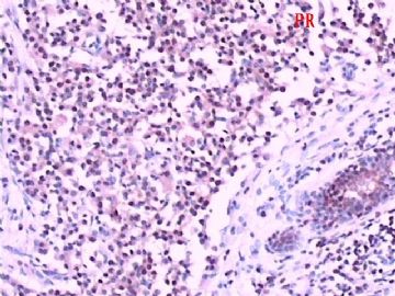
Interesting. CK and ER/PR stains are convencing. Above first four photos may represent DCIS with necrosis, photos 5, 7 may represent invasion. I am not sure why the photos look very strange. Is it the nature of the lesion or caused by fixation (time not enough) or autolosis (keeping fresh tissue for a long time before fixation).
I feel pain to make dx based on these slides.
Only for your reference.

