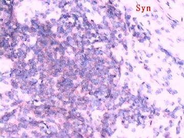
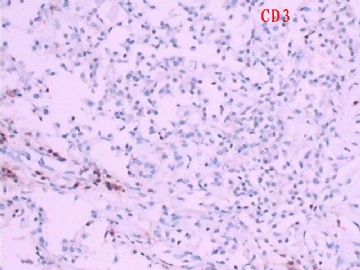
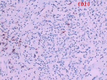
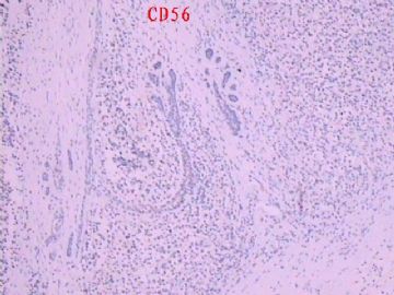
| 图片: | |
|---|---|
| 名称: | |
| 描述: | |
- B1783左乳肿物(罕见病例)
| 姓 名: | ××× | 性别: | 女 | 年龄: | 50 |
| 标本名称: | 左乳腺肿物 | ||||
| 简要病史: | |||||
| 肉眼检查: | 乳腺肿物,直径2cm | ||||

- 把我们应该做的做到最好!
相关帖子
-
liguoxia71 离线
- 帖子:4174
- 粉蓝豆:3122
- 经验:4677
- 注册时间:2007-04-01
- 加关注 | 发消息
-
本帖最后由 于 2009-04-24 23:14:00 编辑
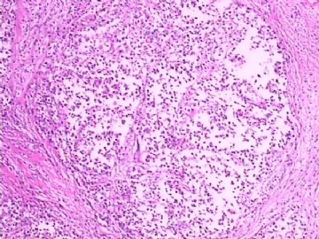
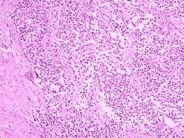
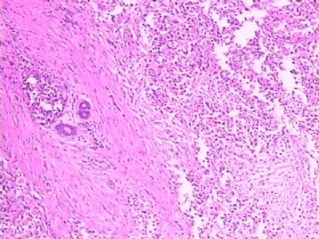
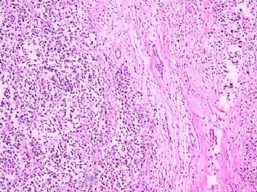
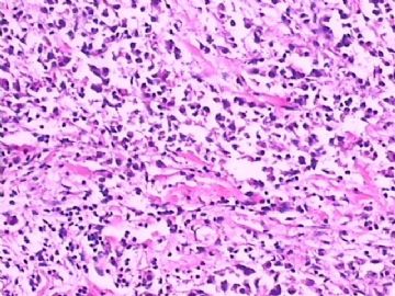
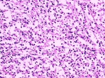
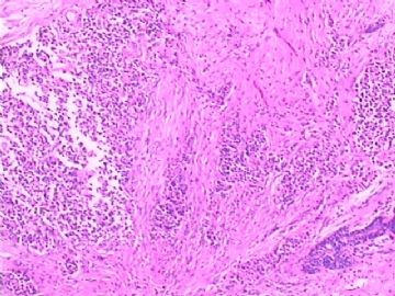
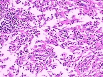
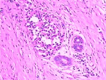
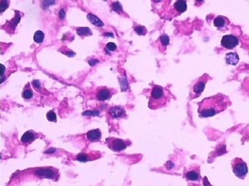
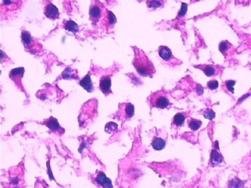
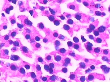
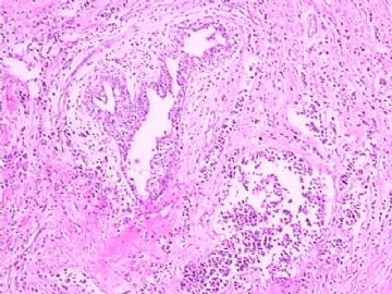
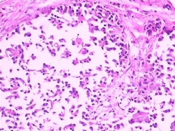
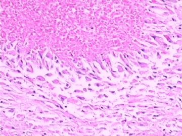
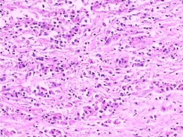
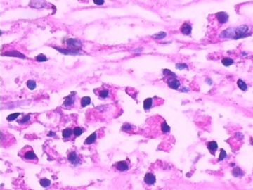
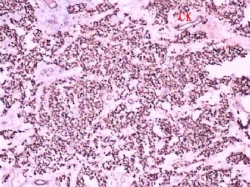
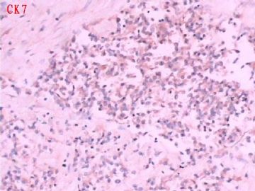
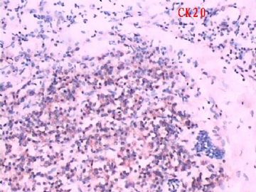
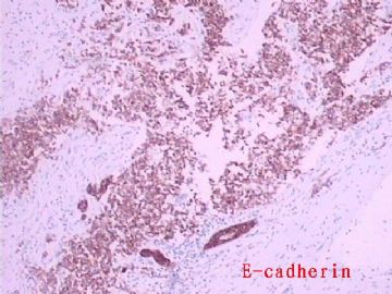
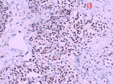
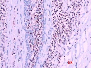
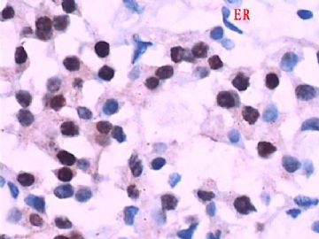
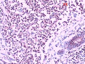
Interesting. CK and ER/PR stains are convencing. Above first four photos may represent DCIS with necrosis, photos 5, 7 may represent invasion. I am not sure why the photos look very strange. Is it the nature of the lesion or caused by fixation (time not enough) or autolosis (keeping fresh tissue for a long time before fixation).
I feel pain to make dx based on these slides.
Only for your reference.
-
liguoxia71 离线
- 帖子:4174
- 粉蓝豆:3122
- 经验:4677
- 注册时间:2007-04-01
- 加关注 | 发消息
-
本帖最后由 于 2009-04-24 23:18:00 编辑
试着 翻译cqzhao老师贴子,请各位指正:
有趣的病例!CK和ER/PR染色可信。头4张图显示了伴坏死的导管原位癌,图5、7显示有浸润。不知道为什么图片look very strange?是病变本身表现还是标本固定时间不充分或标本自溶?靠这些图片做鉴别诊断困难。
仅供参考!
Should be "the photos look very strange".
Dr. liguoxia71
Sorry for my typing error.
Thank you for translation.
cz

- 三人行,必有我师焉,择其善者而从之,其不善者而改之。
Thank for Dr. 楚江渔夫 and Chiang's reasonable explanation.
So this is not a rare case. However this is a good example to demostrate why all 工作流程中的每个细节 are equal important in pathology.
If sugeons did not send the sample to the department of pathology it was the problem of surgeons. If surgeons sent to the dept of pathology on time and people in the pathology dept did not fix the specimen on time it is problem of pathologists or technicians.
In fact this is a big problem if it happens in the USA. Some one needs to fill in an error report to the quality control department in the hospital.
Fixation is a very important issue. It can cause the difficulty for original diagnosis and also can change IHC resutls. Some IHC results can be too important for diagnosis and treatment. In the US CAP requires at least 6 hours' fixation and optimal 8 hours for breast fixation to do the Her2/nue stains.

