共10页/192条首页上一页34567下一页尾页
回复:192 阅读:20192
B1778双侧乳腺血管肉瘤,有近期随访

wfbjwt 离线

帖子:6628
粉蓝豆:163
经验:7418
注册时间:2006-10-08
加关注  |  发消息
楼主 发表于 2009-04-15 17:10|举报|关注(7)
浏览排序[ 顺序 逆序 楼主 支持 精彩 ]  快捷回复
姓    名: ××× 性别:  女 年龄:  28
标本名称:  
简要病史:  
肉眼检查:  
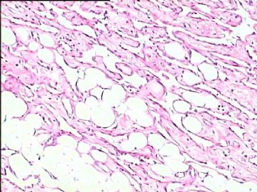
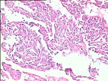
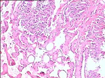
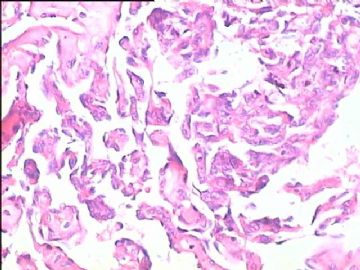
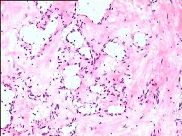
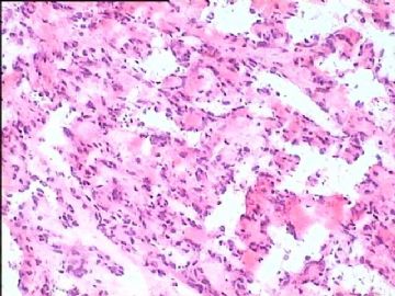
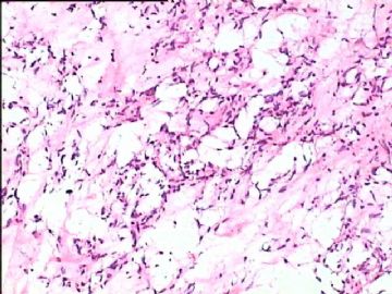
1年前断乳,随后双侧乳腺肿胀,按乳汁潴留、乳腺修复不全保守治疗,2月前左乳切开引流。1-9石蜡切片,10-12冷冻切片。
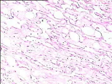
  • 双侧乳腺血管肉瘤,有近期随访图1
    图1
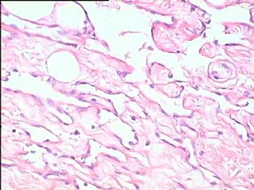
  • 双侧乳腺血管肉瘤,有近期随访图2
    图2
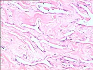
  • 双侧乳腺血管肉瘤,有近期随访图3
    图3
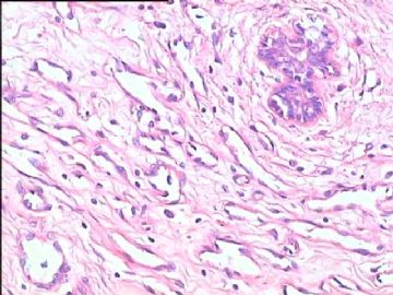
  • 双侧乳腺血管肉瘤,有近期随访图4
    图4
  • 双侧乳腺血管肉瘤,有近期随访图5
    图5
  • 双侧乳腺血管肉瘤,有近期随访图6
    图6
  • 双侧乳腺血管肉瘤,有近期随访图7
    图7
  • 双侧乳腺血管肉瘤,有近期随访图8
    图8
  • 双侧乳腺血管肉瘤,有近期随访图9
    图9
  • 双侧乳腺血管肉瘤,有近期随访图10
    图10
  • 双侧乳腺血管肉瘤,有近期随访图11
    图11
  • 双侧乳腺血管肉瘤,有近期随访图12
    图12
标签:血管肉瘤 PASH 假血管瘤样间质增生 血管瘤
本帖最后由 于 2010-01-29 19:27:00 编辑
1
signature
嫁人就嫁灰太狼,学习要上华夏网。
×参考诊断
双侧乳腺血管肉瘤,高分化

tyl27987 离线

帖子:11
粉蓝豆:1
经验:12
注册时间:2009-03-28
加关注  |  发消息
81 楼    发表于2009-07-07 12:09:00举报|引用
返回顶部 | 快捷回复

 首先考虑良性的,根据有:切片中的乳腺小叶没有被破坏,那些血管样结构内车细胞都不肥胖,异型性不明显,也没有坏死出血等改变。会不会是假血管瘤样纤维组织增生,考虑到妊娠因素,那些看似官腔一样的结构会不会是在妊娠条件下假血管更明显的反应。其次还考虑有没有可能是淋巴管瘤,最后再考虑高分化血管肉瘤。水平有限,大家多指教。期盼最后诊断。

0
回复

chenkf 离线

帖子:230
粉蓝豆:1868
经验:429
注册时间:2007-06-19
加关注  |  发消息
82 楼    发表于2009-07-07 14:41:00举报|引用
返回顶部 | 快捷回复

 学习了,谢谢!

0
回复

wfbjwt 离线

帖子:6628
粉蓝豆:163
经验:7418
注册时间:2006-10-08
加关注  |  发消息
83 楼    发表于2009-07-07 22:20:00举报|引用
返回顶部 | 快捷回复
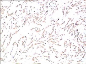
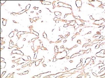
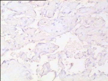
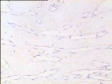
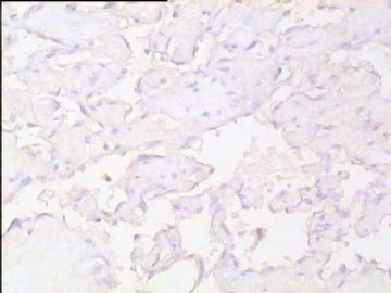
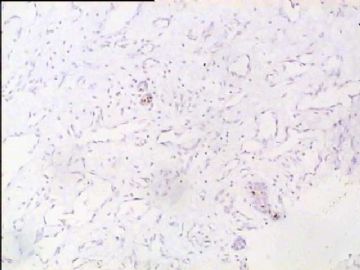
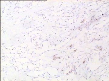
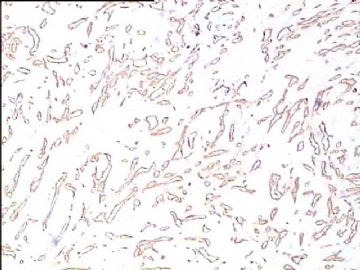
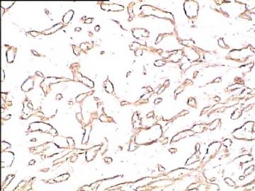
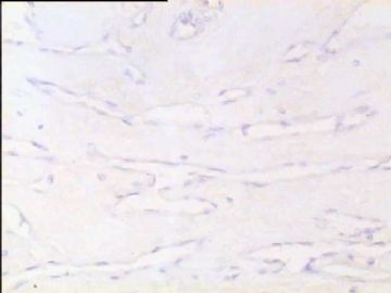
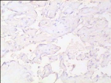
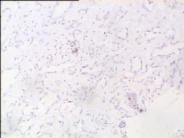
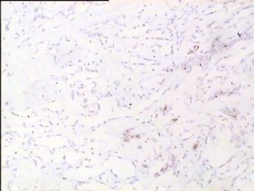
 CD34(+),Ki-67(-),PR(-),萎缩导管PR(+).
0
回复
signature
嫁人就嫁灰太狼,学习要上华夏网。

江边观潮人 离线

帖子:2684
粉蓝豆:524
经验:3338
注册时间:2006-11-12
加关注  |  发消息
84 楼    发表于2009-07-07 22:34:00举报|引用
返回顶部 | 快捷回复
 值得我认真反复学习的病例,谢谢大家!
0
回复
signature

华夏

huocheng_325 离线

帖子:152
粉蓝豆:6
经验:247
注册时间:2009-04-15
加关注  |  发消息
85 楼    发表于2009-07-07 22:40:00举报|引用
返回顶部 | 快捷回复
 考虑1、乳腺血管瘤?2、假血管瘤样乳腺间质增生?(
0
回复
signature
理解万岁。

ynzy1 离线

帖子:187
粉蓝豆:247
经验:844
注册时间:2009-06-08
加关注  |  发消息
86 楼    发表于2009-07-07 22:46:00举报|引用
返回顶部 | 快捷回复
 脉管肿瘤,冰冻不能轻易下血管肉瘤,等石蜡。
0
回复
signature
张银

天天田田 离线

帖子:493
粉蓝豆:5
经验:493
注册时间:2009-05-15
加关注  |  发消息
87 楼    发表于2009-06-21 12:25:00举报|引用
返回顶部 | 快捷回复

感谢赵老师的精彩分析,受益匪浅,受益的不仅仅是专业知识,谢谢,各位老师.

0
回复

junyu_guo 离线

帖子:2
粉蓝豆:1
经验:2
注册时间:2009-06-21
加关注  |  发消息
88 楼    发表于2009-06-21 02:08:00举报|引用
返回顶部 | 快捷回复
 看核分裂定血管肉瘤!
0
回复
signature
多谢关照

klwzfh 离线

帖子:495
粉蓝豆:18
经验:1345
注册时间:2006-11-05
加关注  |  发消息
89 楼    发表于2009-07-10 10:15:00举报|引用
返回顶部 | 快捷回复
 又复习一遍
0
回复

cqzhao 离线

帖子:5518
粉蓝豆:1071
经验:5755
注册时间:2008-09-29
加关注  |  发消息
90 楼    发表于2009-07-10 11:06:00举报|引用
返回顶部 | 快捷回复
以下是引用wfbjwt在2009-7-7 22:20:00的发言:

 CD34(+),Ki-67(-),PR(-),萎缩导管PR(+).

Thank Dr wfbjwt for your information.

What do you mean ki67 negative? Is it deadly negative?

0
回复

wfbjwt 离线

帖子:6628
粉蓝豆:163
经验:7418
注册时间:2006-10-08
加关注  |  发消息
91 楼    发表于2009-07-10 19:19:00举报|引用
返回顶部 | 快捷回复
本帖最后由 于 2009-07-10 19:28:00 编辑  我们Ki-67试剂比较稳定,应是阴性。做的是高分化区域,细胞难以观察,应该做2级的区域就好了。
0
回复
signature
嫁人就嫁灰太狼,学习要上华夏网。

Dr_Lu 离线

帖子:111
粉蓝豆:1018
经验:139
注册时间:2007-12-06
加关注  |  发消息
92 楼    发表于2009-06-22 17:52:00举报|引用
返回顶部 | 快捷回复
  我从头到尾看了这一例的讨论,真的是很有意思。

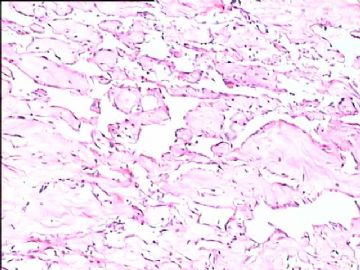
其实乳腺的这种高分化血管肉瘤在会诊和读片会中并不少见,北京市读片会、全国病理读片会多次有相同的病例,06年(也可能是05年?)IAP香港区病理年会时M.D Andson Cancer Center的教授展示过同样一例乳腺高分化血管肉瘤。

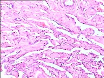
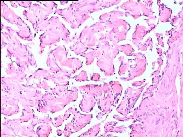
我们之所以不敢下决心诊断,原因是对这种病变的组织学了解不足,大家看一下这个图片,再对照一下软组织病理学(Soft Tissue Tumor,蓝皮的新版的又出来了),软组织中这种看似良性的相互沟通的鹿角状、迷路状相互沟通的血管腔隙,是诊断的特征性病变。以后看到得多了,胆子自然就大了。这种高分化血管肉瘤预后并不好,对治疗反应很差。

有一种情况下,也可出现与上述非常相似的病变,是在血栓机化时,这时决不能诊断为高分化血管肉瘤,一定要慎之再慎!

类似的情况下冰冻直接诊断,风险还是比较大的。如果遇到这种情况,建议跟外科大夫沟通,告诉他们我大概有9成左右的把握,还有一些类似的病变需要鉴别。请他们与病人家属商量,以决定手术是否继续进行。在事先沟通的情况下,一般不会发生后续的医疗纠纷。

总之,冰冻有风险,大家(病理大夫、外科大夫、病人家属)共承担,病理大夫不要逞强,独自承担风险。

0
回复
signature
我的微博:眼镜加显微镜 访问地址:http://weibo.com/pathology

wfbjwt 离线

帖子:6628
粉蓝豆:163
经验:7418
注册时间:2006-10-08
加关注  |  发消息
93 楼    发表于2009-07-12 13:26:00举报|引用
返回顶部 | 快捷回复
本帖最后由 于 2009-07-12 13:30:00 编辑 继续补充资料。包括术前增强CT图片,假体取出时愈合不良组织,及CD34、Ki67、PR各2张。
  • 图1
  • 图2
  • 图3
  • 图4
  • 图5
  • 图6
  • 图7
  • 图8
  • 图9
  • 图10
  • 图11
  • 图12
  • 图13
  • 图14
  • 图15
  • 图16
  • 图17
  • 图18
0
回复
signature
嫁人就嫁灰太狼,学习要上华夏网。

吴梦2008 离线

帖子:44
粉蓝豆:1
经验:44
注册时间:2008-04-15
加关注  |  发消息
94 楼    发表于2009-07-12 15:30:00举报|引用
返回顶部 | 快捷回复
 

 乳腺血管肉瘤

好病例。学习

0
回复

skybobo 离线

帖子:1312
粉蓝豆:154
经验:2232
注册时间:2009-06-22
加关注  |  发消息
95 楼    发表于2009-06-23 22:50:00举报|引用
返回顶部 | 快捷回复
 高分化血管肉瘤
0
回复
signature
病理乃医学之魂

cqzhao 离线

帖子:5518
粉蓝豆:1071
经验:5755
注册时间:2008-09-29
加关注  |  发消息
96 楼    发表于2009-06-24 12:18:00举报|引用
返回顶部 | 快捷回复
本帖最后由 于 2009-06-24 02:13:00 编辑

 Thank Dr. Lu for sharing you oppinion. Looks that you read the true glass slides. Wish you can have more time to come the web to teach more pathology knowledge.

Thanks,

cz

0
回复

kjy7878 离线

帖子:134
粉蓝豆:23
经验:136
注册时间:2008-05-07
加关注  |  发消息
97 楼    发表于2009-06-24 12:19:00举报|引用
返回顶部 | 快捷回复
 学习了。
0
回复

cqzhao 离线

帖子:5518
粉蓝豆:1071
经验:5755
注册时间:2008-09-29
加关注  |  发消息
98 楼    发表于2009-06-24 12:53:00举报|引用
返回顶部 | 快捷回复
 Am J Surg Pathol. 2008 Jun;32(6):943-50. onmouseout="PopUpMenu2_Hide();" href="javascript:PopUpMenu2_Set(Menu18551753);" target="_self">Links

Atypical vascular lesions after surgery and radiation of the breast: a clinicopathologic study of 32 cases analyzing histologic heterogeneity and association with angiosarcoma.

Department of Pathology and Laboratory Medicine, Emory University School of Medicine, Atlanta, GA 30322, USA.

We report the clinicopathologic study of 32 cases of atypical vascular lesions (AVLs) after surgery and radiation of the breast, which were referred to us in consultation over a 17-year period. The patients, all women, ranged in age from 41 to 95 years (mean 61 y). The lesions developed within the radiation field from 1 to 12 years (median 6.0 y) after therapy. They occurred as one (n=18) or more (n=10) flesh-colored papules or erythematous patches/plaques ranging in size from 1 to 60 mm (mean 8.0 mm, median 4.0 mm). Tumors could be divided into 2 histologic types: a lymphatic type (LT) (n=22) and a vascular type (VT) (n=10). LT AVLs consisted predominantly of thin-walled, variably anastomosing lymphatic vessels that were usually confined to the superficial dermis but occasionally extended into the deep dermis and even subcutis. The VT (n=10) typically consisted of small, irregularly dispersed, often blood-filled, pericyte-invested, capillary-sized vessels involving the superficial or deep dermis. VTs were often associated with extravasated erythrocytes, hemosiderin, and a surrounding minor LT component. In 4 cases, endothelial atypia, consisting of nuclear and nucleolar enlargement, was noted. Follow-up of 21 patients with LT AVLs (1 to 106 mo; mean 47 mo) disclosed recurrence/additional lesions in 6, all of whom had additional surgery. Of the 21 patients, 17 are alive without disease, 1 is alive with disease, 1 died of breast carcinoma, 1 died of unknown causes, and 1 showed progressive histologic changes in the AVLs over a period of 5 years resulting in a well-differentiated angiosarcoma. Follow-up in 8 patients with VT AVL (2 to 181 mo; mean 40 mo) disclosed that 6 were alive and well, but 2 of the 4 patients whose lesions displayed endothelial atypia had additional complications. One patient underwent a mastectomy that revealed extensive residual AVL and the second developed a high-grade angiosarcoma after 14 months. We conclude that AVLs encompass a wider spectrum of changes than previously appreciated, ranging from superficial lymphatic proliferations to more complex lymphatic and capillary vascular lesions. There seems to be an association of AVL with angiosarcoma that differs depending on the histologic features, with the VT AVLs having the higher risk. In the 2 patients who developed angiosarcoma, morphologic evidence suggested AVLs to be a precursor rather than simply a risk factor. Future outcome and management studies should take into account these differences.

0
回复

xxl2495 离线

帖子:525
粉蓝豆:73
经验:1018
注册时间:2007-09-26
加关注  |  发消息
99 楼    发表于2009-06-30 23:10:00举报|引用
返回顶部 | 快捷回复

 学习!学习!

好例子!!!

0
回复
signature
当心智平静时,世界也平静。

黄果树 离线

帖子:143
粉蓝豆:335
经验:262
注册时间:2009-02-15
加关注  |  发消息
100 楼    发表于2009-07-01 17:47:00举报|引用
返回顶部 | 快捷回复
  高分化血管肉瘤。学习了!但若碰到了,冷冻切片仍不敢报。
0
回复
回复:192 阅读:20192
共10页/192条首页上一页34567下一页尾页
【免责声明】讨论内容仅作学术交流之用,不作为诊疗依据,由此而引起的法律问题作者及本站不承担任何责任。
快速回复
进入高级回复
您最多可输入10000个汉字,按 "Ctrl" + "Enter" 直接发送
搜索回复/乘电梯 ×
按内容
按会员
乘电梯
合作伙伴
友情链接