| 图片: | |
|---|---|
| 名称: | |
| 描述: | |
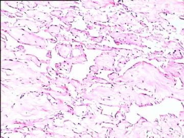
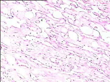
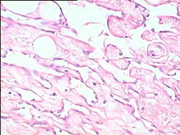
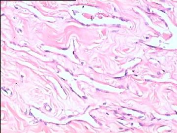
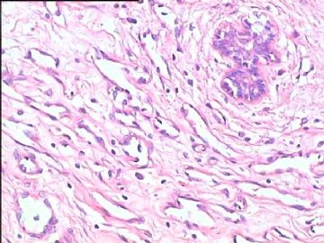
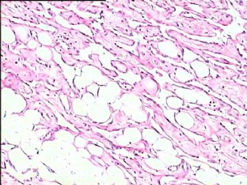
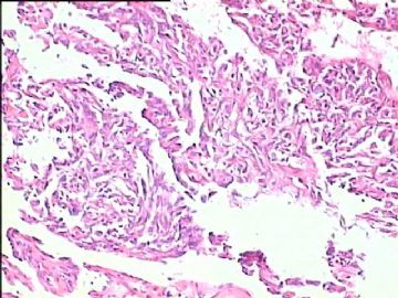
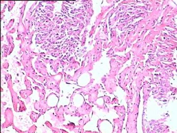
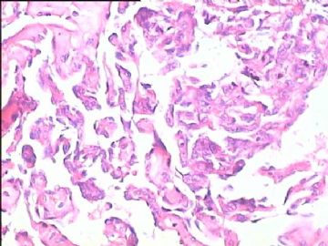
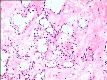
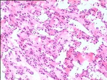
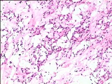
- B1778双侧乳腺血管肉瘤,有近期随访
| 姓 名: | ××× | 性别: | 女 | 年龄: | 28 |
| 标本名称: | |||||
| 简要病史: | |||||
| 肉眼检查: | |||||
-
本帖最后由 于 2010-01-29 19:27:00 编辑

- 嫁人就嫁灰太狼,学习要上华夏网。
相关帖子
- • 乳腺肿物一例,请会诊。
- • 乳腺肿块
- • 右乳包块(男)
-
huisheng97 离线
- 帖子:263
- 粉蓝豆:22
- 经验:285
- 注册时间:2009-02-13
- 加关注 | 发消息
-
susansusan 离线
- 帖子:150
- 粉蓝豆:1
- 经验:270
- 注册时间:2008-11-30
- 加关注 | 发消息
-
zhao_yanfeng 离线
- 帖子:2
- 粉蓝豆:1
- 经验:2
- 注册时间:2009-03-17
- 加关注 | 发消息
-
zhao_yanfeng 离线
- 帖子:2
- 粉蓝豆:1
- 经验:2
- 注册时间:2009-03-17
- 加关注 | 发消息
-
本帖最后由 于 2009-04-25 20:51:00 编辑
Interesting case.
In the USA , most western countries, Asian countries including Japan, India, et al, for breast lesions FNA or core bx is the first initial dx. I know we still do the open biopsy with frozen for breast mass in many hospitals in China. I think it is crazy in term of patient care and difficulty for pathology (frozen fat tissue). We still are using the old method which was not used by most countries for 30 or 40 years. The key is that open biopsy with frozen is not cost effect and is easily to have wrong diagnosis. A lot of patients have segmental mastectomy which may be not necessary. As pathologists we know that it is difficult for us to make dx based on perminant H@E slides for a lot of cases. How can you make dx for some ADH, low grade DCIS, focal high grade DCIS, focal invasive ca, tubular ca vs adenosis, low grade invasive ca vs. sclerosing adenosis, et al.
We have 30, 000/year surgical specimes, half of breast cases and half of gynecologic cases. I work here for three years and never do any breast frozen for original dx. All patients have breast core biopsy before they have excisional, segmental or toal mastectomy.
We some times do frozen for sentinel lymph node. Surgens will decide if the axillary lymph nodes 清扫术 should be done.
We inked margins and cut the segmental or mastectomy specimen to evaluate margin grossly, but not frozen of margins.
-
本帖最后由 于 2009-06-16 12:28:00 编辑
Seems most people think it is angiosarcoma case. This is soft tissue pathology area. Clearly it is not a obvious dx for benign or malignant. I know very little on soft tissue and will think more about this case. Hemangioma is not a very uncommon lesion in breast. I was in breast core biopsy banch in the past week. I had about 90 breast core biopsy cases. I saw two cases of small hemangioma.
I have a quesion for your guys especially for 楼主。Obviously the differential dx for this case includes low grade angiosarcoma and benign hemangioma. What is your criteria for diangosis of angiosarcoma for this case?
Just for learning purpose. Thanks, cz
我很吃惊这么多人诊断血管肉瘤。我基本不同意该诊断,倾向良性。无核异性,无核分裂,细胞非常小且一致。请问你们诊断血管肉瘤的依据。同意赵大夫意见。血管肉瘤基本不能冰冻切片诊断。
本病例考虑良性血管瘤,pseudoangiomatous stromal hyperplasia (PASH), PASH 细胞ER阳性,a low grade vascular lesion is also in the differential。
我在以前的贴上看到有人说:乳房血管瘤都是恶性,这是不对的,我看到很多乳房良性血管瘤,相反恶行较少。

