| 图片: | |
|---|---|
| 名称: | |
| 描述: | |
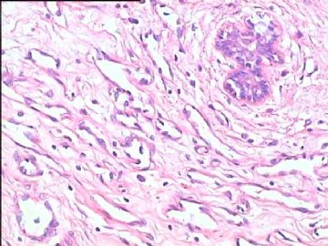
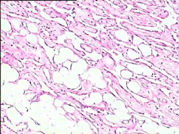
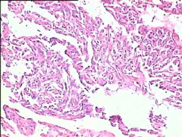
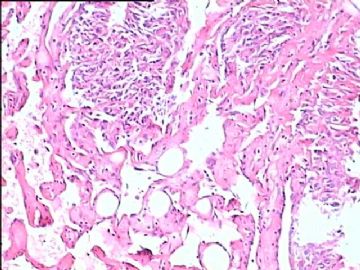
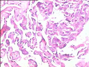
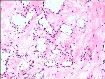
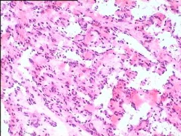
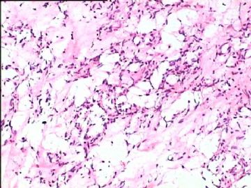
- B1778双侧乳腺血管肉瘤,有近期随访
| 姓 名: | ××× | 性别: | 女 | 年龄: | 28 |
| 标本名称: | |||||
| 简要病史: | |||||
| 肉眼检查: | |||||
-
本帖最后由 于 2010-01-29 19:27:00 编辑

- 嫁人就嫁灰太狼,学习要上华夏网。
相关帖子
- • 乳腺肿物一例,请会诊。
- • 乳腺肿块
- • 右乳包块(男)
| 以下是引用klwzfh在2009-5-18 11:26:00的发言:
 |
I feel sad when I saw the discussion like this. I spent a lot of time on this case and tried to let people know how to make the dx for some difficult cases. It may be a sarcoma case. I cannot make the dx or rule out the dx based on the photos. Also I pasted the comment from many soft tissue expert pathologists including world well known soft tissue pathologists. Of cause experts can make the wrong dx also.
血管肉瘤,印象中记得 Gee老师讲过,乳腺血管瘤极少,多数都是恶性,即使形态良善。按此知道精神一定诊断:血管肉瘤
Pathologists cannot make dx based on this kind of logic thinking.
-
hongpinguo 离线
- 帖子:92
- 粉蓝豆:142
- 经验:109
- 注册时间:2009-01-16
- 加关注 | 发消息
乳腺假血管瘤样间质增生:
Vuitch等于1986年首先描述了假血管瘤样间质增生(pseudoangiomatous stroma hyperplasia,PASH)作为乳腺一种镜下或肉眼的独立性病变,主要表现为乳腺间质的疤痕样纤维化,其内有裂隙状假血管样间隙。PASH的病因还不十分清楚,可能与激素水平紊乱有关。
临床表现 大多数见于绝经前的妇女,14~67岁,平均37岁。通常在一侧乳腺发现无痛性、界限清楚的硬块。少数病例局部增厚,界限不清,乳腺弥漫肿大者罕见。
眼观 病变区呈结节状,大小1.2~12 cm,平均6 cm,质地硬,多数界限比较清楚,但没有包膜,切面橡皮样,灰白色,有时可见小囊腔,没有出血和坏死。
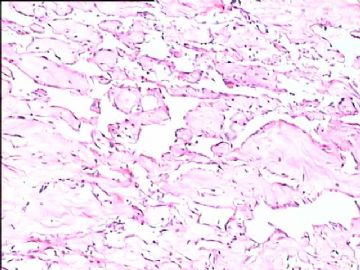
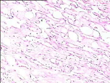
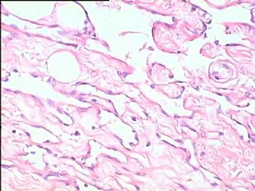
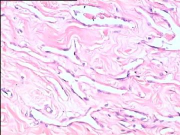
镜检 小叶间有广泛的疤痕样纤维组织增生,伴有不规则的裂隙样间隙(类似毛细血管),间隙腔是空的,不含红细胞,其壁被覆内皮样梭形细胞(电镜和免疫组化证实为纤维母细胞)(图4),梭形细胞可有明显增生和轻度异型性,但无核分裂,也不呈丛状生长。病变常围绕乳腺小叶,也可长进小叶内,小叶结构一般存在,但其间隙增宽。病变周围常存在纤维囊性病变、纤维腺瘤、男性乳腺发育、硬化性腺病或正常乳腺。[/font][/color][/size]

- 广州金域病理
-
本帖最后由 于 2009-05-01 21:07:00 编辑
反复看图,先写下自己的思路:
图1-6:迷路样血管样腔隙,被覆细胞无异型性,间质细胞稀疏,胶原化,穿插在小叶间(图5)。图9-12细胞增生,密度增高。一些圆形腔是脂肪细胞?看不清。所有图中腔内均未见红细胞。
结合病史:患者28岁,断乳一年,双侧。考虑1、乳腺血管瘤?2、假血管瘤样乳腺间质增生?(pseudoangiomatous stroma hyperplasia,PASH)。
不过以上多为一侧发生,中青年。此例双侧,会不会与断乳后引起的乳汁储留修复不全有关。

- 广州金域病理
-
本帖最后由 于 2009-07-27 07:23:00 编辑
Copy a few sentences from Rosen's breast pathology book:
For primary breast angiosarcoma:
Total mastectomy is the recommended primary surgical therapy.
Axillary dissection is not indicaterd because metastases rarely involv these lymph nodes.
The role of radiation has not been determined.
Following surgery, systemic adjuvant chemitherapy may ne offered, but the effectiveness of this treatment remains uncertain.

