| 图片: | |
|---|---|
| 名称: | |
| 描述: | |
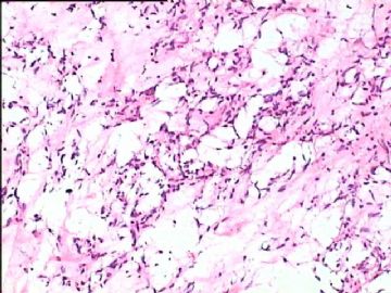
- B1778双侧乳腺血管肉瘤,有近期随访
| 姓 名: | ××× | 性别: | 女 | 年龄: | 28 |
| 标本名称: | |||||
| 简要病史: | |||||
| 肉眼检查: | |||||
-
本帖最后由 于 2010-01-29 19:27:00 编辑

- 嫁人就嫁灰太狼,学习要上华夏网。
相关帖子
- • 乳腺肿物一例,请会诊。
- • 乳腺肿块
- • 右乳包块(男)
-
本帖最后由 于 2010-01-30 11:17:00 编辑
Thank Dr. wfbjwt for the follow up. Now no doubt it is angiosarcoma because patient has recurrent and also distant metastasis.
Now we can go back to review the case.
1. Clearly cytology is very bland.
2. Why is it a malignant tumor?
多发结节状生长,无包膜,周围组织界限不清 and tumor size. These features seem important for evaluation of the breast vascular lesions.
I once asked several experts to review the photos and got different oppinions. I think there are two reasons. Firstly this is a difficult case. More important fact to affect the evaluation is that they only saw a few photos, but not true all slides and gross specimen.
In fact after I got different oppinions from expert pathologist I mentioned above, I sent a few photos to other top soft tissue pathologists in the US or in the world. Dr. Christopher Fletcher(Harvard Medical School) thought it was a angiosarcoma, but another pathologist was not sure the diagnosis.
Anyway it is sad for the young women. However it is a good case for us to learn.
Thanks wfbjwt again.
-
本帖最后由 于 2010-01-30 20:24:00 编辑
This is a challenge and debatable case in the begining. It is complete case from frozen to final dx to follow up result. It was reviewed and consulted in several hospitals in China. The photos were reviewed by several internatonal famous soft tissue pathologists for their oppinions. Nice case for learning.
It is difficult to evaluate some challenge cases based on a few photos online.
大家在网站的討论建议仅供参考。特别是对基层医院的病理医生,无论别人怎样建议,你在签发病理报告时必须真正了解和同意这个诊断,即使是上级医院的专家会诊意见,because it is your own case.
总 置顶 few days for more people to learn the lesson.
We all should thank Drwfbjwt for providing the case.
cz
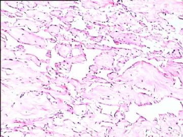
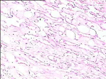
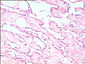
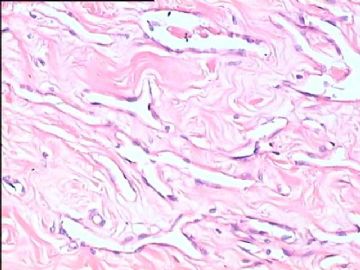
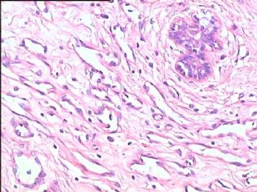
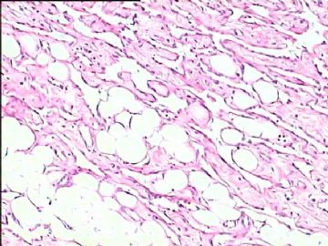
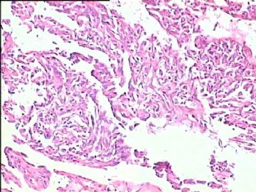
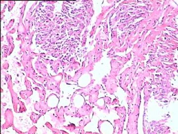
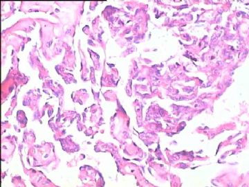
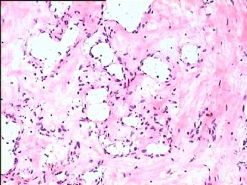
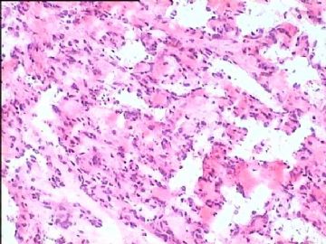
所有图片均显示没有细胞异形、没有不典型性、没有坏死、没有浸润,窦样间隙内也看不到红细胞,其中一张图见病变围绕小叶但小叶结构正常,血管肉瘤的诊断能成立吗?乳腺血管肉瘤可以分化好,但必须具有血管肉瘤的特征。
本例形态特征符合乳腺假血管瘤样间质增生。其形成原因可能与哺乳后的内分泌紊乱有关。镜下特征有复杂的血窦样间隙,内衬内皮细胞样梭形细胞,缺乏异形性和核分裂象,间质为透明变性的增生纤维组织,低倍镜易误认为低级别血管肉瘤,但其细胞学特征和生长方式缺乏恶性证据可资鉴别。免疫组化可表达CD34、vim、actin、calponin,不表达CD31、S-100、EMA、CD68、CK、8因子等。



 我以为是谁误操作呢呵呵
