| 图片: | |
|---|---|
| 名称: | |
| 描述: | |
- B1777乳腺囊肿壁
-
lfl001200546 离线
- 帖子:2808
- 粉蓝豆:40
- 经验:2808
- 注册时间:2007-02-14
- 加关注 | 发消息
This is true difficult case especially based a few photos. Differential diagnosis includes benign mucocele-like lesion and mucinous carcinoma.
Focal cystic wall lined with benign looking epithelial cells with associated myoepithelial cells, hemosiderin-macrophages (suggestive rupture of the cysts), detached clusters of epithelium within the mucin (often present in mucinous carcinoma, but they have benign looking in this case). Basically my feeling is that this is mucocele-like benign lesion. However I cannot make the diagnosis only based on the above photos in true case. I have to read all true glass slides with microscopy for diagnosis.
So above just for your reference.
-
liguoxia71 离线
- 帖子:4174
- 粉蓝豆:3122
- 经验:4677
- 注册时间:2007-04-01
- 加关注 | 发消息
-
haiyan7981 离线
- 帖子:11
- 粉蓝豆:1
- 经验:11
- 注册时间:2009-03-27
- 加关注 | 发消息

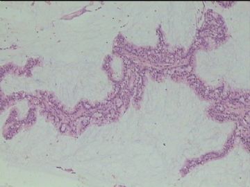
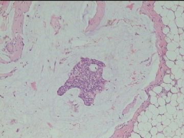
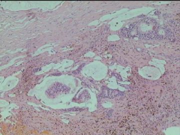
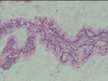
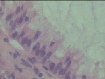
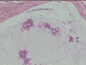



 考虑乳腺粘液性囊肿病,但需做免疫组化除外粘液癌。


