| 图片: | |
|---|---|
| 名称: | |
| 描述: | |
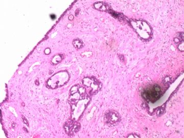
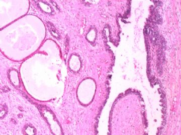
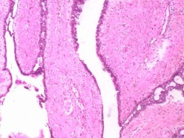
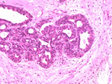
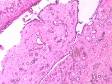
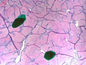
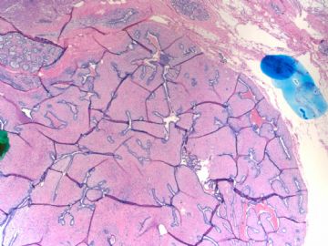
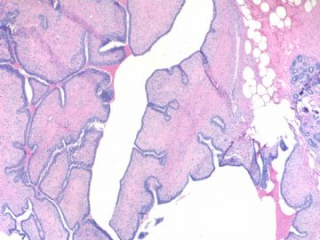
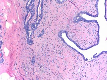
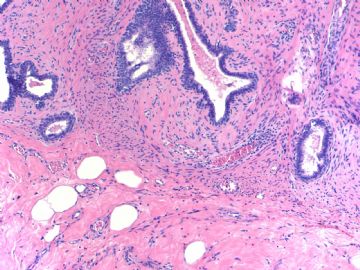
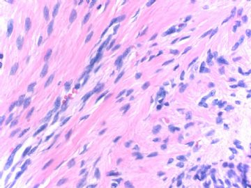
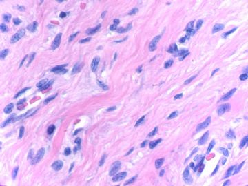
- B1770Breast fibroepithelial lesions (cqz 15)
| 姓 名: | ××× | 性别: | 年龄: | ||
| 标本名称: | |||||
| 简要病史: | |||||
| 肉眼检查: | |||||
I notoced there are some cases of fibroepithelial lesions (biphasic tumor)in our website. I send this case to open the discussion.
Fibroepithelial tumors including fibroadenoma (FA) and phyllodes tumors (PT). The definition and classification of PT are not very agreeable among pathologists. However, they are most common classified as bengin, borderline, or malignant suggested by WHO. Some pathologists use terms of benign, low grade maligant and high grade malignant. These two systems are very similar, borderlin equal to low grade malignant.
Hope you can give your diagnosis and mention why. I assume people may have different oppinions. It is ok. We just discuss the case.
相关帖子
Well well done. Excellent summary. Thank 笃 (du) 行者 . Hope more people can involve the website more actively. We can think the web as a free stage. Every one can come here to share, show, or discuss the cases. When you prepare a summary you need to review the book and you learn in the same time. Often I need to review the books or papers to answer the questions. By the way I learn a lot from the book and from all of your discussion.
Thank 笃行者 again. People whoever read your summary can know the main points quickly.
cz
| 以下是引用杨宝军在2009-4-17 23:23:00的发言: 请问老师们如果第2例是原发的诊断叶状肿瘤的依据?? |
借此话题,复习一下叶状肿瘤的诊断,希望对年轻医生有所帮助。
关于良性、交界性和恶性叶状肿瘤的诊断,WHO列出了三者鉴别诊断的参考指标:
观察指标 良性 交界性 恶性
间质细胞增生 少量 少量 明显
间质细胞多形性 轻度 中度 明显
核分裂像数 少(<3) 中(3~5) 多(>10个/10HPF)
边缘情况 界限清楚 中 浸润性生长
间质分布 均匀 不均匀 明显过度增生
异源性分化 罕见 罕见 常见
免疫组化对于良恶性叶状肿瘤诊断也有一定的帮助:(本人对此没有经验)
良性叶状肿瘤:bcl-2 + 、β-catenin + 、CD117 -
恶性叶状肿瘤:bcl-2 -/+ 、β-catenin - 、CD117 +
虽然大家都在参考这个标准,可是具体到某一个病例则诊断可能有很大的分歧,那就看你怎样掌握它使用它了
对于叶状肿瘤的诊断,出现困难的情况我想,一个是良性叶状肿瘤和纤维腺瘤的鉴别;一个是恶性叶状肿瘤与其他肉瘤和梭形细胞癌的鉴别。
良性叶状肿瘤,它相对于纤维腺瘤有以下特点:
1、年龄相对较大(一般40岁以上,25岁以下罕见)。
2、肿块近期增长速度较快,体积相对较大(一般>5cm)(肿物体积大小并不特别重要)
3、大体检查:表面常有突起,切面具有可见的裂隙而呈分叶状,有时呈菜花样。
4、低倍镜下可见较大的裂隙,呈扩大化的管内型生长方式。
5、间质细胞相对丰富、呈轻度的增生活跃、轻度的间质细胞核多形性和相对的间质过度增生。
但是,上述任何单独一项都不具特异性,都是相对性的。如果将上述情况结合起来,则大多数情况下可以将两者鉴别开来。
尽管如此,对于某些病例,如在富于细胞的纤维腺瘤和良性叶状肿瘤之间做出鉴别是非常困难的,有的甚至是不可能的,至今还没有一个非常可靠和被普遍接受的鉴别依据。
虽说间质细胞丰富是叶状肿瘤最重要的形态特征,但有时也可以不丰富;而在幼年型纤维腺瘤(富于细胞性纤维腺瘤),其间质却可以很丰富。此时年龄因素很重要。另外,幼年型纤维腺瘤导管上皮常明显增生。
另外对于某些鉴别困难的病例,下列情况可能有帮助:
间质内有明显化生(如脂肪、骨、软骨、横纹肌等);
上皮有较多鳞状化生;
——多考虑叶状肿瘤
病变内出现大汗腺化生
——多考虑纤维腺瘤
想到那里说到哪里,没有系统性,也不知是否有意义、观点是否正确。如有错误,请大家,尤其是cqzhao老师给予指正,谢谢。

- 博学之,审问之,慎思之,明辨之,笃行之。
-
zhang197510 离线
- 帖子:409
- 粉蓝豆:2971
- 经验:448
- 注册时间:2009-03-22
- 加关注 | 发消息
Dr. Yang,
You are right. I cannot make the dx of phyllodes if it were the primary lesion for the second case. The dx of recurrent phyllodes was based on the pt's phyllodes history, the same location, and irregular border. The criteria are made by people, they often cannot be used for all cases. Individual case should be considered or treated individually in practical pathologic diagnosis.
| 以下是引用cqzhao在2009-4-16 2:24:00的发言:
Thank 笃行者 . I forget how to call the word 笃 even though you told me before. Please teacher me again. I will remember this time. cz |
呵呵,其实这个字以前我也不会读,查字典才知道“笃”读作"du",与“赌、堵”同音,“忠实,一心一意”的意思。
很高兴在网上认识赵老师,并且很荣幸在网上一睹赵老师的容颜。

- 博学之,审问之,慎思之,明辨之,笃行之。


QIE[D1B}RQ)O}M07SHB6.jpg)
5QJ4CV(($V`Z05V09.jpg)
