| 图片: | |
|---|---|
| 名称: | |
| 描述: | |
- B1770Breast fibroepithelial lesions (cqz 15)
| 姓 名: | ××× | 性别: | 年龄: | ||
| 标本名称: | |||||
| 简要病史: | |||||
| 肉眼检查: | |||||
I notoced there are some cases of fibroepithelial lesions (biphasic tumor)in our website. I send this case to open the discussion.
Fibroepithelial tumors including fibroadenoma (FA) and phyllodes tumors (PT). The definition and classification of PT are not very agreeable among pathologists. However, they are most common classified as bengin, borderline, or malignant suggested by WHO. Some pathologists use terms of benign, low grade maligant and high grade malignant. These two systems are very similar, borderlin equal to low grade malignant.
Hope you can give your diagnosis and mention why. I assume people may have different oppinions. It is ok. We just discuss the case.
相关帖子
Well well done. Excellent summary. Thank 笃 (du) 行者 . Hope more people can involve the website more actively. We can think the web as a free stage. Every one can come here to share, show, or discuss the cases. When you prepare a summary you need to review the book and you learn in the same time. Often I need to review the books or papers to answer the questions. By the way I learn a lot from the book and from all of your discussion.
Thank 笃行者 again. People whoever read your summary can know the main points quickly.
cz
Dr. Yang,
You are right. I cannot make the dx of phyllodes if it were the primary lesion for the second case. The dx of recurrent phyllodes was based on the pt's phyllodes history, the same location, and irregular border. The criteria are made by people, they often cannot be used for all cases. Individual case should be considered or treated individually in practical pathologic diagnosis.
Dr. Qianxun,
Thank you very much for your comment. For phyllodes tumors we count mitosis very carefully. Personally I never use ki67 stain for diagnosis of phyllodes. Could you describe the proliferative index more details for distinguishing benign from borderlin phyllodes? Thanks, cz
-
About case 2: Patient had history of phyllodes tumor several years ago (outside hospital). So this is local recurrence of benign phyllodes. It is difficult to predict the prognosis for phyllodes. The major concern is local recurrence. Distant metastases are rare.Complete excision with wide free margin is the main treatment. One large review study indicated the chance of local recurrence was 21%, 46%, and 65% for the patients with benign, borderline, and malignant phyllodes.
-
本帖最后由 于 2009-03-14 11:05:00 编辑
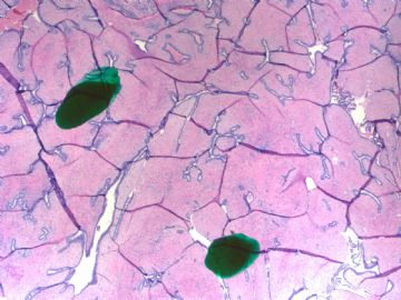
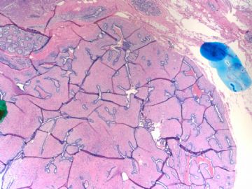
I am showing you another fibroepithelial lesion.
Case 2:
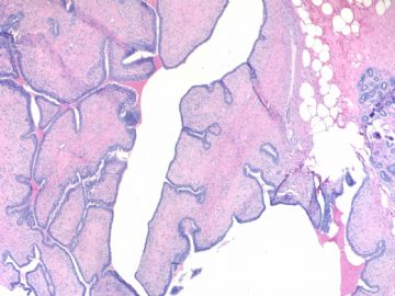
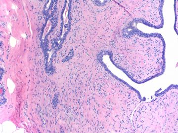
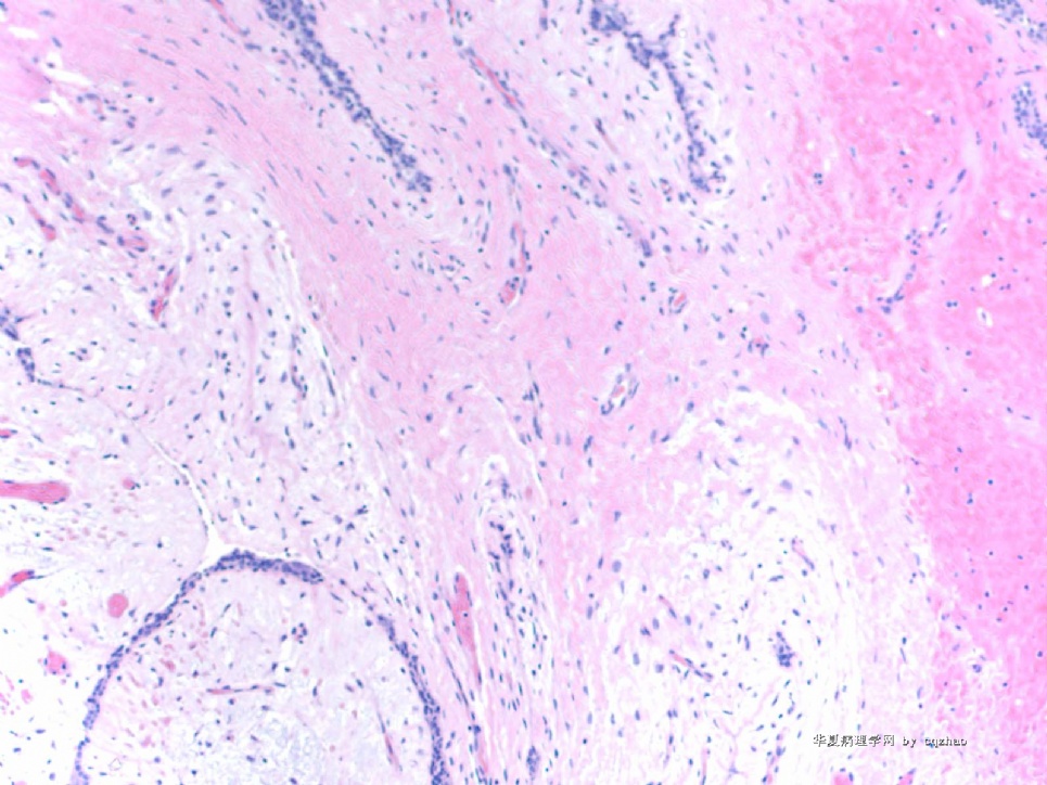
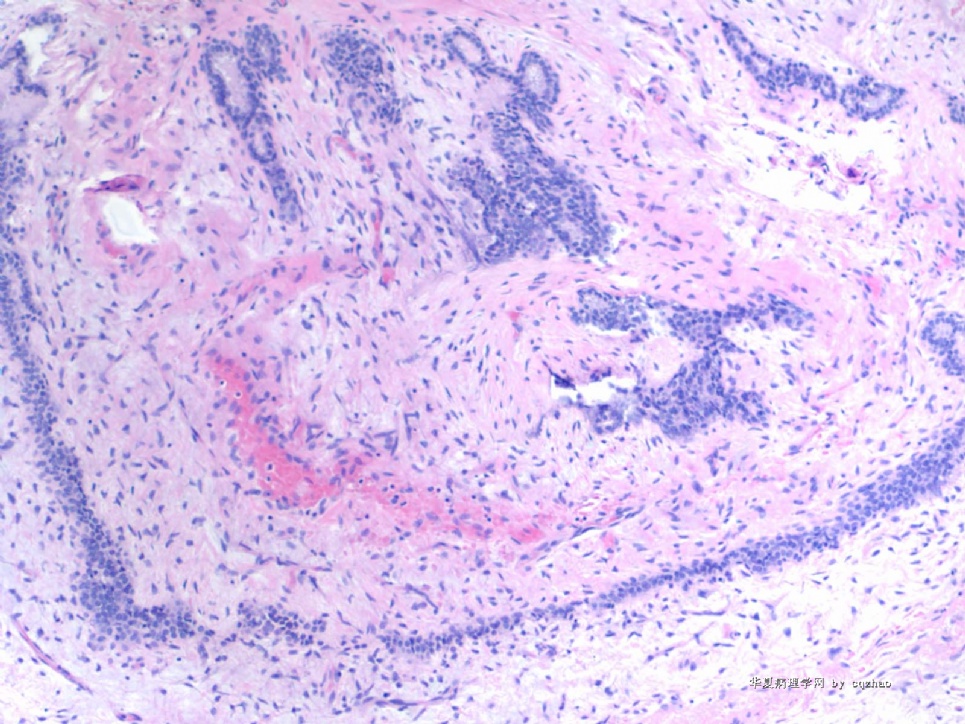
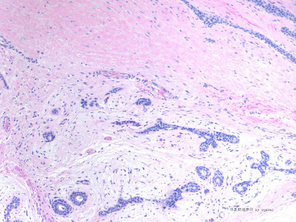
50 y/f right breast lesion 1.7 cm. excisionaly biopsy specimen.
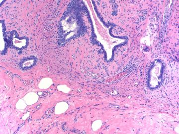
Fig 1. 20x
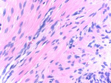
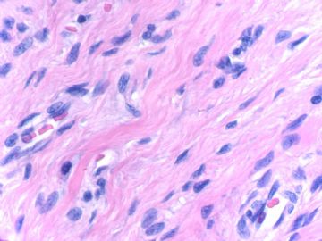
Other fig 100x
No mitosis is noticed.
Your diagnosis?
名称:图1
描述:图1

名称:图2
描述:图2

名称:图3
描述:图3
名称:图4
描述:图4
名称:图5
描述:图5
名称:图6
描述:图6
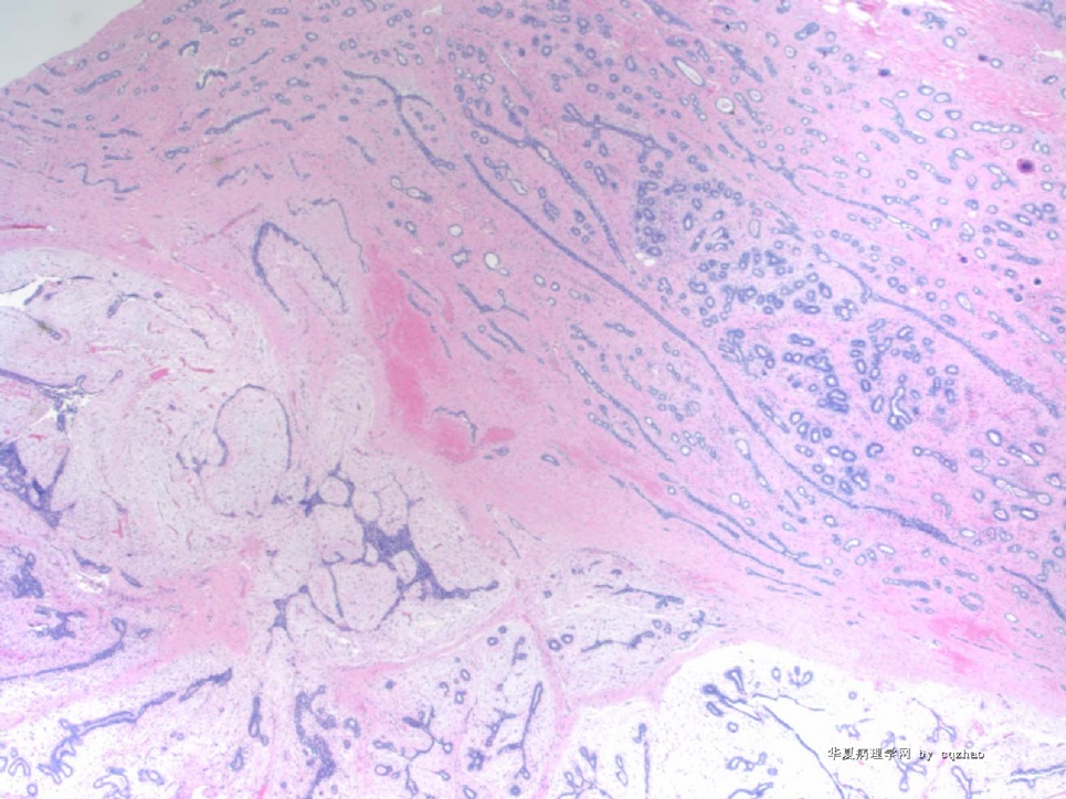
Most of people here have an agreenment for this case. The tumor demonstrates leaf-like structures, dense stroma with a little heterologeneous distribution and mild cytologic atypia, and a little increased cellularity, pushing margin with focal irregularity. Mitotic firures are rare (0-1/10 high power fields). Clearly it should be a phyllodes tumor, but not fibroadenoma, even though the tumor is small. Considering all the facts I diagnosed benign phyllodes finally.
Welcome you to share your different oppinion.
