| 图片: | |
|---|---|
| 名称: | |
| 描述: | |
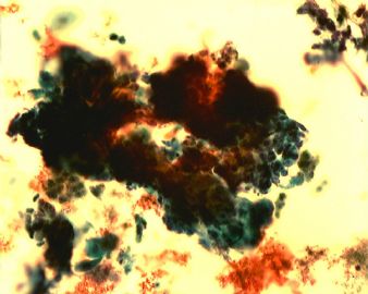
- 41y宫颈液基
Thank you very much. This really is an unique case. Plus 全子 had histologic follow-up result. Basaloid squamous cell carcinoma is a high grade malignant carcinoma. Do not confused with adenoid basal carcinoma, which is low grade tumor.
Pap test with follow-up result is key for learning.
-
本帖最后由 于 2009-03-12 19:52:00 编辑
感谢赵老师,毕竟是高手,把我打的小埋伏都点出来了
回赵老师:
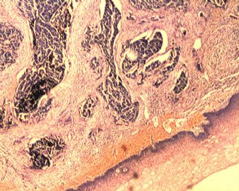
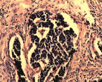
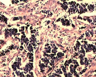
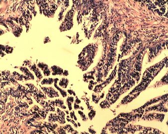
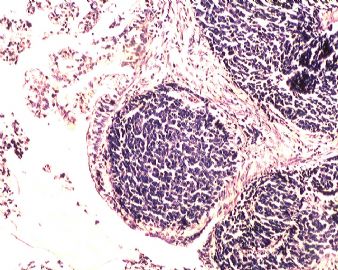
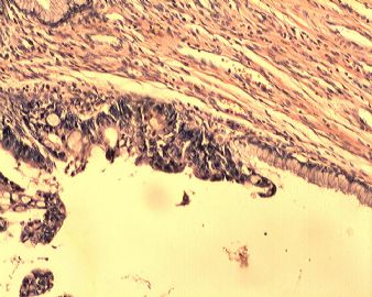
1.HE显示的结构中,腺癌是明确的,本例腺样结构和团块状结构则显示了腺癌的特点,这一点大家都注意到了
2.那些小细胞是什么,我认为是基底细胞样鳞癌伴有神经内分泌分化,因为这个本来是细胞贴,没有详细描述,您说的几个类型的确是需要鉴别的。
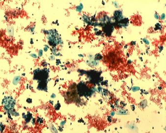
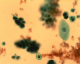
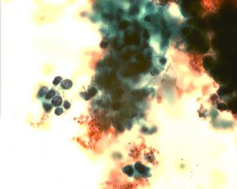
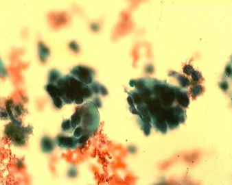
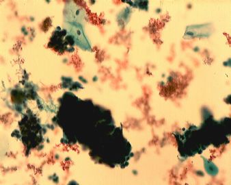
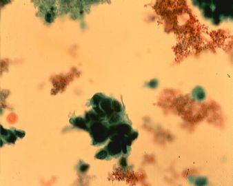
3.再回过头去看细胞学表现,那些散在的小簇状细胞也正是基底细胞样癌的细胞学表现了,其鉴别散在的腺癌细胞的要点在于,细胞的形态并不是腺上皮的柱状细胞,而是小的底层细胞表现呈小圆形、立方状,本例不是角化形鳞癌所以并未显示其角化特点,而且簇状的排列比较松散。值得注意的是,那些小细胞也正如不少网友看到的是否有一些“小纤毛”,这个是不是神经内分泌分化的癌的细胞学特点有待于进一步考证。
4.宫颈CIN的诊断中其实是经常(至少不少见)可以看见宫颈腺体的异型变化的,这一点也提请大家不要忽略,这也是本例拿出来的意义所在
一点愚见,望批评指正
Just notice 全子's case. Very good case. Now I can say what I want to because i know the histologic result already. Difficult to tell it is squamous lesion or squamous lesion with glnadular involvement or glandular lesion based on cytology. fell it is a bad lesion with with bleeding background. In fact this is an excellent case. We can see some clusters of glandular cells with marked atypia, also we can appreciate some samll clusters of small cells with the cells in Fig 2, 3, 5. We can appreciate two types of cells clearly in Fig 3. They are adenocarcinoma in histologic fig 4 and 6. What are these samll cells in histology fig 2,3, 5? Small malignant cells show very dark nuclei with increase N/c ratio. The growth paterns like glandular cells, but cytologic features like small cell carcinoma, neuroendocrine carcinoma, adenoid basal ca, basaloid squamous carcinoma. They are not like classic squamous cell carcinoma.
全子: thank for sharing the interesting case with cyto and histologic results.
Could you tell us what are these small cells? thanks, cz



 学习了
学习了 


