| 图片: | |
|---|---|
| 名称: | |
| 描述: | |
- B1527宫腔刮出物
-
wangdingding 离线
- 帖子:1474
- 粉蓝豆:98
- 经验:6042
- 注册时间:2006-10-19
- 加关注 | 发消息
| 姓 名: | ××× | 性别: | 女 | 年龄: | 45 |
| 标本名称: | |||||
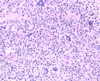
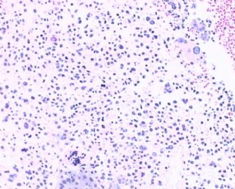
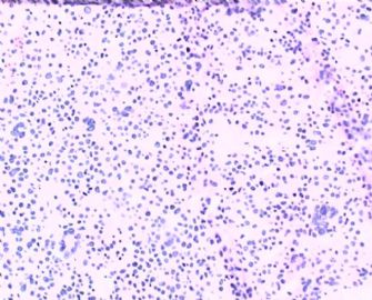
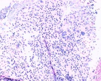
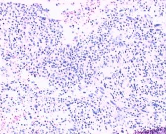
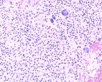
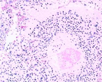
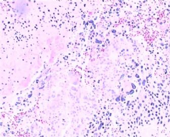
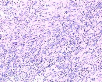
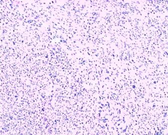
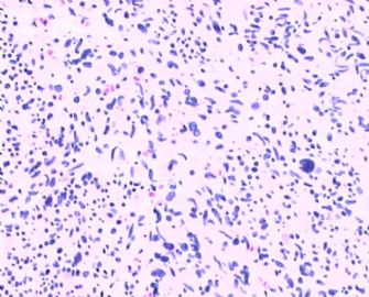
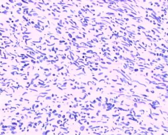
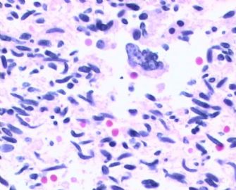
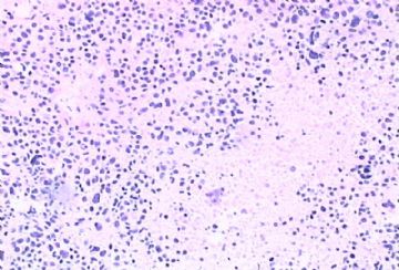
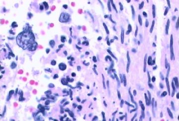
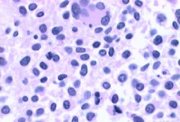
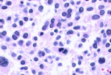
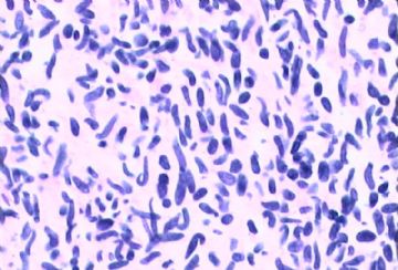
| 简要病史: | 阴道不规则出血6天,B超发现子宫后壁肌瘤,术中探宫深10cm,刮出组织约10g. | ||||
| 肉眼检查: | 暗红碎组织一堆:3X2X1.5CM. | ||||
相关帖子
- • 来一例简单罕见的(有诊断)
- • 是肉瘤吗?
- • 子宫内膜,复杂性增生?癌?(有大体结果了)
- • 子宫肿物(透明细胞平滑肌瘤?)
- • 子宫肌壁间肿物
- • 子宫腔内占位。
- • 子宫肿瘤
- • 子宫肌层浸润性癌
- • 8 个子宫上皮肿瘤病例-扫描图片
- • 子宫平滑肌肿瘤?间质肉瘤?
-
huisheng97 离线
- 帖子:263
- 粉蓝豆:22
- 经验:285
- 注册时间:2009-02-13
- 加关注 | 发消息
-
huisheng97 离线
- 帖子:263
- 粉蓝豆:22
- 经验:285
- 注册时间:2009-02-13
- 加关注 | 发消息
| 以下是引用LIU_AIJUN在2009-4-30 23:57:00的发言: 请关注坏死、核分裂像、异型性和瘤体与正常肌壁的关系,综合评判定良恶。 |
Agree with above.
1. Floor 10 and 14 photos from hystectomy specimen look like leiomyomas with infarction (not coagulate tumor necrosis). I cannot appreciate mitosis and atypia from these photos.
2. Floor One EMC specimen: tumor cells demonstrate marked cytologic atypia. Need to count the mitosis carefully from the true glass slides. It seems there are a few mitoses based on the photos.
Cell types: It is strange that both epithelial markers and vimentin were negative. Also smooth muscle markers were negative.
Try more different smooth muscle markers. Some pleomorphic leiomyosarcoma can be smooth muscle markers negative, but at least some markers may be focally or weekly positive.
Try CD10 to rule out stromal tumor.
Try Inhibin and calretinin, CD99 stains to rule out uterin tumor resembling ovarian sex cord tumor. The chance is very low.
If smooth muscle tumor is confirmed, marked cytologic atypia plus mitotic figures >10/10hpf, no tumor cell necrosis in photos in floor one, the tumor is leomyosarcoma. Otherwise it is atypical leiomyoma with low risk of recurrence (or STUMP).
It is impossible to evaluate these kinds of complicated cases based on a few photos inline.
Something likes 纸上谈兵。 ha, ha.
