| 图片: | |
|---|---|
| 名称: | |
| 描述: | |
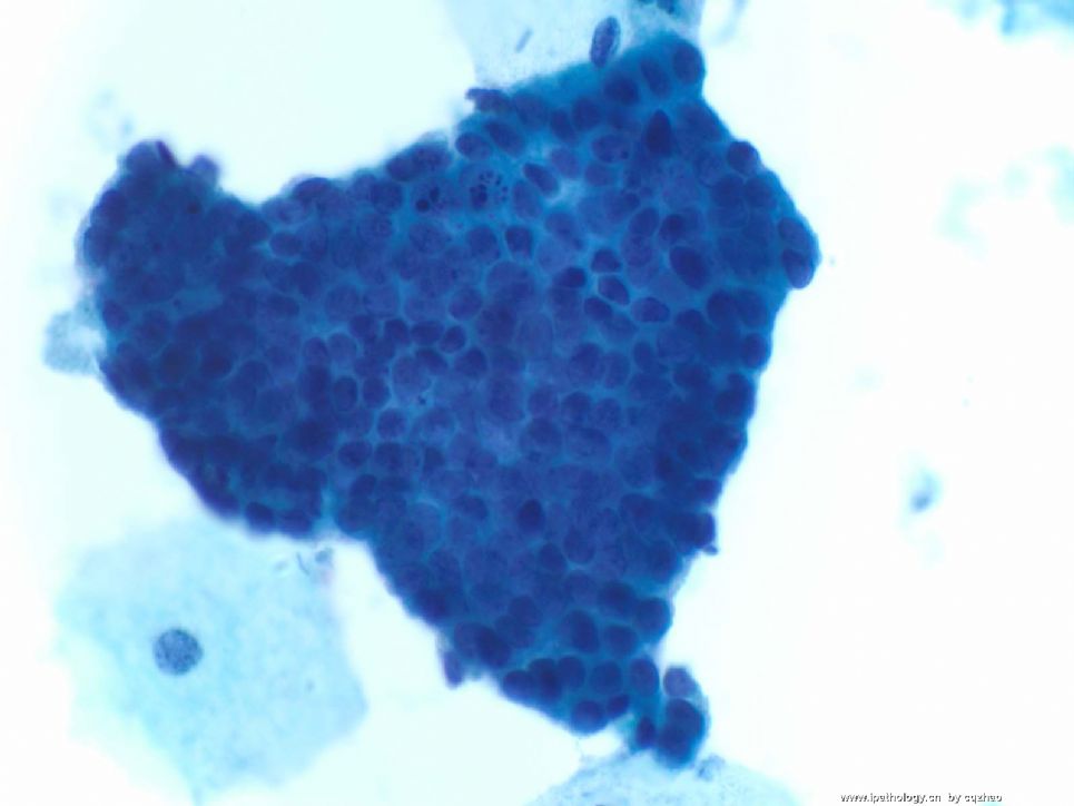
- an easy case for you-Pap test (cqz 3)
-
I sent similar photos from this case to another chines pathology website. It seems that more people write down their interpretation. Now it is the new year eve here and is the first day of 2009 already in China. Wish more people can join in the discussion more actively. I am going to help to prepare the mid night family party. Every one has a wonderful 2009.
|
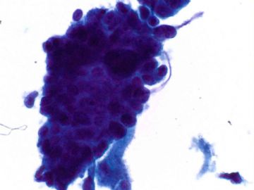
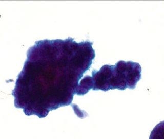
我感觉两幅都没有太大的问题。第一幅象子宫内膜腺上皮细胞团,第二幅图象宫颈内膜腺上皮细胞团。如果是我的病例,但是她是一位50岁的人,建议病人一月后复查! |
The two clusters are similar. Both are hyperchromatic crowded groups (HCG) of cells with big, dark nuclei. Are you sure they are ok? generally do not need to repeat Pap in one month until the Pap smear is unsatisfactory. |
| 以下是引用月新在2008-12-30 11:03:00的发言:
译赵老师:two different interpretation above. 以上有两种不同的解释,One is likely from endometrial origin and anther one is from endocervical origin. 一种认为是子宫内膜来源,另一种认为是宫颈内膜来源。What is other pathologists' oppinion? 听一下病理医生们的意见? 我感觉两幅都没有太大的问题。第一幅象子宫内膜腺上皮细胞团,第二幅图象宫颈内膜腺上皮细胞团。如果是我的病例,但是她是一位50岁的人,建议病人一月后复查! |
译赵老师:two interpretation above. 上边两幅图,One is likely from endometrial origin and anther one is from endocervical origin. 一幅象子宫内膜,另一幅象宫颈内膜。What is other pathologists' oppinion? 听一下病理医生们的意见?
我感觉两幅都没有太大的问题。第一幅象子宫内膜腺上皮细胞团,第二幅图象宫颈内膜腺上皮细胞团。如果是我的病例,但是她是一位50岁的人,建议病人一月后复查!
赵老师:真是不好意思,最近忙没有好好学习,没想到这个帖子这么热闹,而且我还被点名了,汗!仔细看了赵老师和月新老师、望月的分析,受益匪浅,体会最深的是细胞块真是好东西!
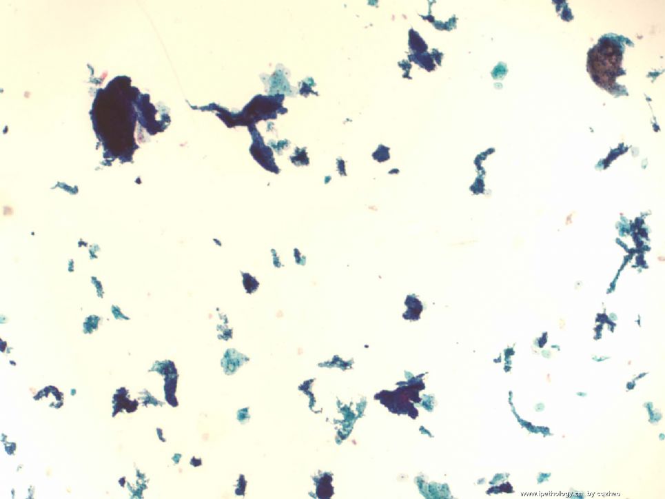
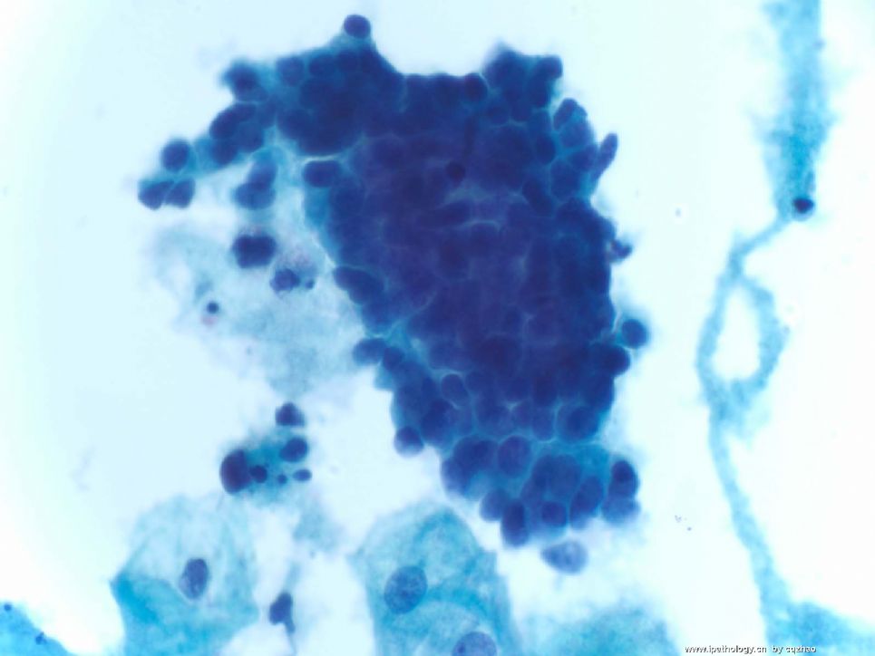
我来看看81楼的病例,成团的腺上皮,我仍然认为它是来源于子宫内膜,只是和上例不同,异型性没有那么大,没有乳头状结构,现在来猜倾向宫内膜腺癌。当然就看两张图片就发评论是需要勇气的(虽然赵老师肯定给出了细胞的典型表现),如果要发报告,我会更加谨慎一点,会了解病史、B超以及妇科检查的情况,发出的报告可能也就是AGC,建议做分段诊刮
也让赵老师教教我,看是不是朽木啦
-
本帖最后由 于 2008-12-28 20:20:00 编辑
| 以下是引用cqzhao在2008-12-24 12:20:00的发言:
Cytomorphology of Papillary Serous Adenocarcinoma n Differential diagnosis: 1. Endometrioid carcinoma: difficult in Pap test 2. Reactive benign endocervical cells: this is the money for pathologists 3 Reactive mesothelial cells (in pelvic washing) n clusters, flat sheets or single cells n Cytoplasm is dense & distinct n Multinucleation n Nuclei chromatin vary from bland to hyperchromatic n Nucleoli-small to prominent |
大致翻译如下:
浆液性乳头状腺癌细胞形态学
鉴别诊断:
1 、子宫内膜样癌:宫颈细胞学诊断很难。
2 、反应性的良性子宫颈细胞:对于病理学家是财富(money)?
3、反应性的间皮细胞(腹水中)
成群、平铺或单个的细胞
细胞质浓密和独特
多核
核染色质从淡染到深染各不相同
核仁:小到明显。

- 广州金域病理
-
本帖最后由 于 2008-12-28 20:17:00 编辑
| 以下是引用cqzhao在2008-12-24 12:13:00的发言:
Cytomorphology of Papillary Serous Adenocarcinoma (Pap) n Malignant cells are isolated or arranged in clusters.
n Nuclei are enlarged and demonstrate variation in size.
n There is nuclear hyperchromasia with coarsely textured chromatin and prominent nucleoli.
n Cytoplasm may be scant but is often abundantly vacuolated.
n Psammoma bodies may be present. |
大致翻译如下:
乳头状浆液性腺癌细胞形态学(巴氏涂片)
恶性细胞单个散在或成簇排列。
细胞核增大,核大小变化较大。
核深染、染色质粗糙、明显的核仁。
细胞质可很少,但常含大量空泡。
可出现沙砾体。

- 广州金域病理
