共3页/59条首页上一页123下一页尾页
回复:150 阅读:16173
an easy case for you-Pap test (cqz 3)

cqzhao 离线

帖子:5518
粉蓝豆:1071
经验:5755
注册时间:2008-09-29
加关注  |  发消息
楼主 发表于 2008-12-12 11:42|举报|关注(0)
浏览排序[ 顺序 逆序 楼主 支持 精彩 ]  快捷回复

Pap smears are too difficult, especially for photos in computer. Give you an easy one for fun.

A women aged over 50

How will you sign out the case?

  • an easy case for you-Pap test  (cqz 3)图1
    图1
  • an easy case for you-Pap test  (cqz 3)图2
    图2
标签:
0
添加参考诊断
×参考诊断
  

cqzhao 离线

帖子:5518
粉蓝豆:1071
经验:5755
注册时间:2008-09-29
加关注  |  发消息
41 楼    发表于2008-12-19 10:41:00举报|引用
返回顶部 | 快捷回复
 Recommend one paper from a very good Chinese pathologist- Fan Lin

Cancer. 2007 Apr 25;111(2):74-82.

Immunohistochemical detection of p16INK4a in liquid-based cytology specimens on cell block sections.

Liu H, Shi J, Wilkerson M, Huang Y, Meschter S, Dupree W, Lin F.

Department of Pathology, Lehigh Valley Hospital, Allentown, Pennsylvania, USA.

BACKGROUND: Colposcopy biopsy procedure is a standard recommendation for atypical squamous cell cannot exclude high-grade lesion (ASC-H) in abnormal Papanicolaou smears. p16 (p16INK4a), a cell cycle regulator, has been shown to be overexpressed in squamous dysplasia. To further improve the diagnostic accuracy of the ASC-H Papanicolaou smear and to reduce unnecessary procedures, the authors evaluated the utility of immunodetection of p16 in liquid-based cytology specimens on cell blocks. METHODS: Seventy-five liquid-based (SurePath; TriPath Imaging, Inc. Burlington, NC) cytology specimens were prepared for cell blocks. Three groups (G1, G2, and G3) of cases were included: G1 comprised 44 cases of ASC-H; G2, 14 cases of high-grade dysplasia; and G3, 17 negative/reactive cases. All cases in G1 were confirmed by cervical biopsy or Digene Hybrid Capture 2 (Digene, Gaithersburg, Md) human papilloma virus (HPV) testing. Immunodetection for p16 was performed on cell blocks. RESULTS: In G1, 26 of 44 (59%) cases showed squamous dysplasia, with 14 high-grade squamous intraepithelial lesion (HSIL) cases. Twenty-two of 28 (79%) p16-positive cases were confirmed by surgical biopsy or HPV testing, with a diagnostic sensitivity of 85%, specificity of 67%, positive predictive value (PPV) of 79%, and negative predictive value (NPV) of 75%. Four cases with false-negative staining for p16 were identified. All 28 cases of HSIL (14 from G1 and 14 from G2) were positive for p16. CONCLUSIONS: 1) p16 is a sensitive marker to confirm the diagnosis of ASC-H on a cell block; 2) Multiple unstained slides with adequate cellularity can be obtained from each cell block; and 3) Additional markers can be used to further increase diagnostic sensitivity and specificity. (c) 2007 American Cancer Society.

0
回复

cqzhao 离线

帖子:5518
粉蓝豆:1071
经验:5755
注册时间:2008-09-29
加关注  |  发消息
42 楼    发表于2008-12-18 22:05:00举报|引用
返回顶部 | 快捷回复
以下是引用小荷在2008-12-18 21:41:00的发言:

 深深谢意!收获多多!

请教一个问题:“无论什么年龄段出现了非典型的的子宫内膜腺上皮细胞都应该做活检,.如果刮宫标本正常还应该再做阴道镜,所以非典型的子宫内膜细胞是一定要报出来。”

如果是非典型腺细胞,不能明确是子宫内膜,还是宫颈管,需要活检吗?!

如果能明确是非典型宫颈管腺细胞,需要活检吗?

You are right:

AGC, endometrial: endometrial and endocervical sampling. need to do colposcopy if no endometrial lesion noted.


All other AGC, (AGC, NOS; AGC, endocervical):

Colposcopy with endocervical sampling, high risk HPV testing. In additon, endometrial sampling is needed for patients are >35 year, or patients have risk for endometrial lesions ---family history, overweight et al.


So AGC is critical call. You have to be think over and try to not overcall or mis-call. A big challenge for all pathologists. I did AGC research for many years and reviewed more than 1000 AGC cases with follow up. I still feel struggle for most cases. Ha, ha. We all need learning from the practice.


It seems my online talk about AGC was not very effective for your guys. Sorry about this. In fact pathologists cannot learn pathology from few talks. Importance is that we learn from our practice and mistakes.


0
回复

cqzhao 离线

帖子:5518
粉蓝豆:1071
经验:5755
注册时间:2008-09-29
加关注  |  发消息
43 楼    发表于2008-12-18 12:43:00举报|引用
返回顶部 | 快捷回复
以下是引用月新在2008-12-18 11:23:00的发言:

 To  天山望月 and 月新 :

It does mean u answer is wrong. 并非说你们的回答是错的,但是不完整。It is true most serous carcinomas are from endometrium, but some can arise from endocervix. 虽然大部分浆液性癌起源于子宫内膜,但是也有部分起源于子宫颈,Just wonder you think you can tell the difference between cervical papillary serous carcinoma and endometrial papillary serous carcioma. 你肯定认为你可以鉴别宫颈或子宫内膜的乳头状浆液性癌,In fact it is very difficult. Endometrioid carcinoma, clear cell carcinoma can arise from endometrium and endocervix. 实事上,子宫内膜样癌,透明细胞癌都可以起源于子宫内膜,也可以起源于子宫颈,Often it is difficult to tell the origins even though you try to use IHC in the biopsy specimens.认清起源是非常困难的,就是用免疫组化也没有用。


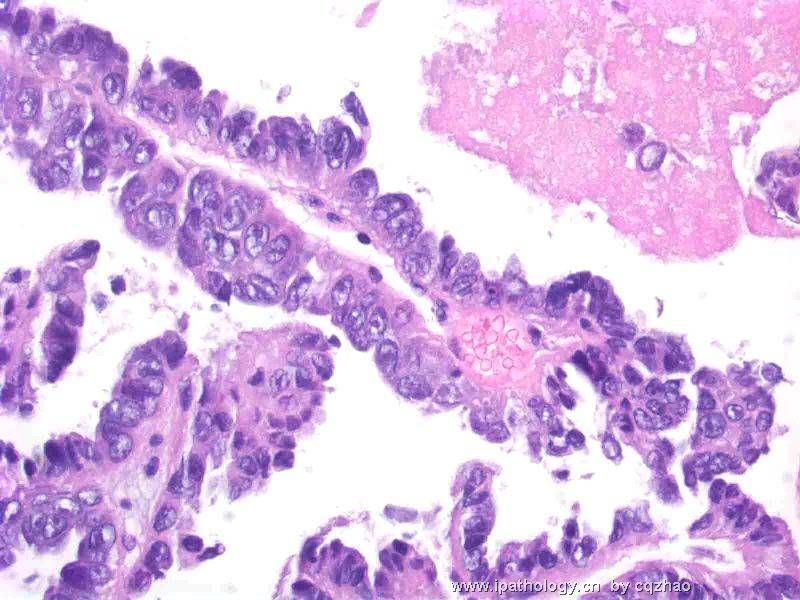
感谢赵老师给我上了一课,一幅图,引出这么多的思维差距,虽然我可以感觉到这是一个恶性的腺上皮细胞团,但是如何进一步工作,如何发病理报告又是不同。看到了宫颈涂片中有上述的异形上皮细胞团,如果光说话让临床医生做这做那,自已不再做实际工作,不行,赵老师用剩余的TCT液体做成细胞块,一下子解决了大问题,把肿瘤的组织形态弄到手了,但是如何描述这样的形态,如何写报告又是一个思维落差。比如是乳突状浆液性腺癌,是子宫颈,还是子宫内膜,大包大揽,肯定说子宫内膜,当然子宫内膜多见,但是宫颈完全可以有。病理报告错一个字,意谓着不完整不科学,也就是完全错误。这一例给我的经验。应该象赵老师这样,做事有缜密性,自已应该做工作一定要做完,思维有多样性,考虑问题复杂,报告有原则性,不能说过头话,原则无论如何不能偏离。这次举一反三。大有收获。

0
回复

cqzhao 离线

帖子:5518
粉蓝豆:1071
经验:5755
注册时间:2008-09-29
加关注  |  发消息
44 楼    发表于2008-12-18 02:00:00举报|引用
返回顶部 | 快捷回复
 For above question I would like to pick C and F. Pay attention to "what are....".
0
回复

cqzhao 离线

帖子:5518
粉蓝豆:1071
经验:5755
注册时间:2008-09-29
加关注  |  发消息
45 楼    发表于2008-12-18 01:55:00举报|引用
返回顶部 | 快捷回复
本帖最后由 于 2008-12-18 01:58:00 编辑

 To  天山望月 and 月新 :

It does mean u answer is wrong. It is true most serous carcinomas are from endometrium, but some can arise from endocervix. Just wonder you think you can tell the difference between cervical papillary serous carcinoma and endometrial papillary serous carcioma. In fact it is very difficult. Endometrioid carcinoma, clear cell carcinoma can arise from endometrium and endocervix. Often it is difficult to tell the origins even though you try to use IHC in the biopsy specimens.

0
回复

cqzhao 离线

帖子:5518
粉蓝豆:1071
经验:5755
注册时间:2008-09-29
加关注  |  发消息
46 楼    发表于2008-12-18 01:49:00举报|引用
返回顶部 | 快捷回复
以下是引用天山望月在2008-12-17 13:28:00的发言:

以下是引用cqzhao在2008-12-17 2:26:00的发言:

以下是引用天山望月在2008-12-16 23:37:00的发言:

 此例细胞块图片非常漂亮,乳头结构典型,乳头状癌。

Do u know from where?

Yes,i know that is from some residual fluid left in the lab .

So important!  hehe


I mean this tumor is from cervical or endometrial origin.
0
回复

cqzhao 离线

帖子:5518
粉蓝豆:1071
经验:5755
注册时间:2008-09-29
加关注  |  发消息
47 楼    发表于2008-12-17 12:46:00举报|引用
返回顶部 | 快捷回复
 

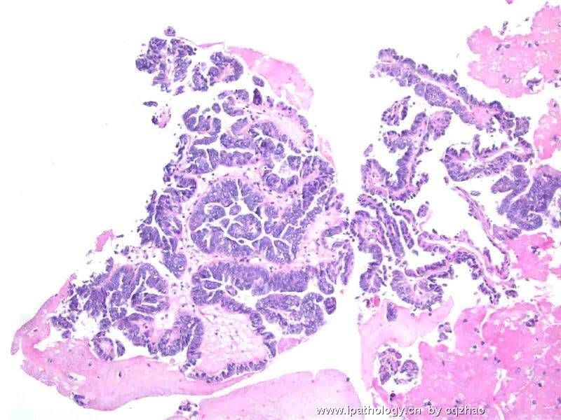
What are the likely diagnosis?

A. Endometrial Endometroid adenocarcinoma

B. Malignant Mixed Mullerian Tumor

C. Endometrial Papillary Serous Adenocarcinoma

D. Reactive Mesothelial Cells

E. Malignant Mesothelioma

F. Endocervical Papillary serous adenocarcinoma
0
回复

cqzhao 离线

帖子:5518
粉蓝豆:1071
经验:5755
注册时间:2008-09-29
加关注  |  发消息
48 楼    发表于2008-12-17 02:26:00举报|引用
返回顶部 | 快捷回复
以下是引用天山望月在2008-12-16 23:37:00的发言:

 此例细胞块图片非常漂亮,乳头结构典型,乳头状癌。

Do u know from where?
0
回复

cqzhao 离线

帖子:5518
粉蓝豆:1071
经验:5755
注册时间:2008-09-29
加关注  |  发消息
49 楼    发表于2008-12-16 22:03:00举报|引用
返回顶部 | 快捷回复
 Now how will you release your pap report?
0
回复

cqzhao 离线

帖子:5518
粉蓝豆:1071
经验:5755
注册时间:2008-09-29
加关注  |  发消息
50 楼    发表于2008-12-16 12:55:00举报|引用
返回顶部 | 快捷回复
本帖最后由 于 2008-12-16 12:58:00 编辑  Based on 天山望月 's order, cell block from residual fluid was prepared in the cyto lab. H&E slide from cell block (low and high power). Now what do you think?

名称:图1
描述:图1

名称:图2
描述:图2
0
回复

cqzhao 离线

帖子:5518
粉蓝豆:1071
经验:5755
注册时间:2008-09-29
加关注  |  发消息
51 楼    发表于2008-12-15 23:06:00举报|引用
返回顶部 | 快捷回复
 I will send you more follow up for this case
0
回复

cqzhao 离线

帖子:5518
粉蓝豆:1071
经验:5755
注册时间:2008-09-29
加关注  |  发消息
52 楼    发表于2008-12-15 23:05:00举报|引用
返回顶部 | 快捷回复
以下是引用法师在2008-12-15 19:38:00的发言:

 1、如果是宫颈管内膜细胞,除非是腺癌,因为正常的宫颈管内膜细胞一般不会呈乳头状,如果是腺癌,又感觉差了点什么。

2、如果是子宫内膜细胞,也除非是癌,正常的子宫内膜细胞没这么大,但也觉得差了什么,子宫内膜癌形成的乳头细胞核之间或多或少有间隙,这个好像很紧凑。

幸好我没遇到,万一真遇到了,就真的糊涂了,就报个非典型腺细胞,无法明确来源,然后再慢慢查去。

You are great based on u name (.法师). You must know every thing. In fact your interpretation is excellent. In true practice you are not wrong if you report as AGC or .... However, it will be wrong if you report benign reactive change. I think every one here can recongnize these clusters of glandular cells are atypical at least. This is why I said it is a very easy case. Remember Pap test is a screening test. Do not need to push yourself soo hard to make a definite diagnosis.
0
回复

cqzhao 离线

帖子:5518
粉蓝豆:1071
经验:5755
注册时间:2008-09-29
加关注  |  发消息
53 楼    发表于2008-12-14 22:19:00举报|引用
返回顶部 | 快捷回复
以下是引用天山望月在2008-12-14 20:15:00的发言:

以下是引用cqzhao在2008-12-14 3:11:00的发言:

 If it were your case above and you know there was some residual fluid left in the lab (10 ml fluid totally), what would you like to do?

回复赵老师:

1、在做TCT时,我喜欢每次将剩余的液体,再离心沉淀备用,如果液基图片有可疑细胞,或制片欠佳,再将备用液制片或做细胞块,再染色观察或做免疫组化等。

2、常与临床沟通,只要有异常细胞,都要和临床联系,询问病史和其它相关检查,并建议做相关的检查,如宫颈外观情况、B超示内膜情况、既往史等。可建议分段清宫、宫颈活检、查HPV、查内分泌激素等。


Excellent. This is what I hope to see. I know our Chinese pathologists are smart.
0
回复

cqzhao 离线

帖子:5518
粉蓝豆:1071
经验:5755
注册时间:2008-09-29
加关注  |  发消息
54 楼    发表于2008-12-14 03:13:00举报|引用
返回顶部 | 快捷回复
 Above all of your analyses are very good. I appreciate your study atitute. Just hope you guys can open your mind and think more.
0
回复

cqzhao 离线

帖子:5518
粉蓝豆:1071
经验:5755
注册时间:2008-09-29
加关注  |  发消息
55 楼    发表于2008-12-14 03:11:00举报|引用
返回顶部 | 快捷回复
 If it were your case above and you know there was some residual fluid left in the lab (10 ml fluid totally), what would you like to do?
0
回复

cqzhao 离线

帖子:5518
粉蓝豆:1071
经验:5755
注册时间:2008-09-29
加关注  |  发消息
56 楼    发表于2008-12-13 12:57:00举报|引用
返回顶部 | 快捷回复

 It is not uncommon to see the normal endometrial cells in women age >45 even the LMP more than 12 days.

We should report the presence of endometrial cells if women 40 y or above. Then we need to mention the EM cells are consistent with LMP, or > 12 days after LMP or do not know the LMP of the patient.

Normal EM cells in women <40, do not need to report  regardless of LMP

Normal EM cells in women 40 or above: Clinical correlation.

Atypical EM cells: should have endometrial and endocervical sampling for all ages. If no endometrial pathology identified, colposcopy should be performed. So atypical EM cells are critical report.

My discussion is no relation to the interpretation of my above case (I do not mean which of your answers is right or wrong). So continue to guess.

0
回复

cqzhao 离线

帖子:5518
粉蓝豆:1071
经验:5755
注册时间:2008-09-29
加关注  |  发消息
57 楼    发表于2008-12-13 12:06:00举报|引用
返回顶部 | 快捷回复

Thank 月新 for your good translation

Normal EM cells in women 40 or above: Clinical correlation. 大于40岁以上的人宫颈涂片中出现正常的子宫内膜细胞. In this situation we can write the comment: Clinical correlation is suggested. It means that clinical physicians should consider patient's clinical stituation to decide how to handle the case. Basically I give the ball to the patient's physicians.


i did not say above answers are right or wrong. Just give a resonable interpretation based on your knowledge.

0
回复

cqzhao 离线

帖子:5518
粉蓝豆:1071
经验:5755
注册时间:2008-09-29
加关注  |  发消息
58 楼    发表于2008-12-12 20:28:00举报|引用
返回顶部 | 快捷回复
以下是引用月新在2008-12-12 17:01:00的发言:

子宫内膜腺上皮细胞团。

normal or abnormal?
0
回复

cqzhao 离线

帖子:5518
粉蓝豆:1071
经验:5755
注册时间:2008-09-29
加关注  |  发消息
59 楼    发表于2008-12-12 12:52:00举报|引用
返回顶部 | 快捷回复
以下是引用wy1992在2008-12-12 11:50:00的发言:

 非典型腺上皮,建议随访.染色深可能是染片的原因

Friend, I am interested to know where you learn above?

You want your AGC women have Pap follow up. Hope you are joking. Ha, ha. 

0
回复
回复:150 阅读:16173
共3页/59条首页上一页123下一页尾页
【免责声明】讨论内容仅作学术交流之用,不作为诊疗依据,由此而引起的法律问题作者及本站不承担任何责任。
快速回复
进入高级回复
您最多可输入10000个汉字,按 "Ctrl" + "Enter" 直接发送
搜索回复/乘电梯 ×
按内容
按会员
乘电梯
合作伙伴
友情链接