| 图片: | |
|---|---|
| 名称: | |
| 描述: | |
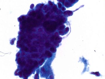
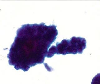
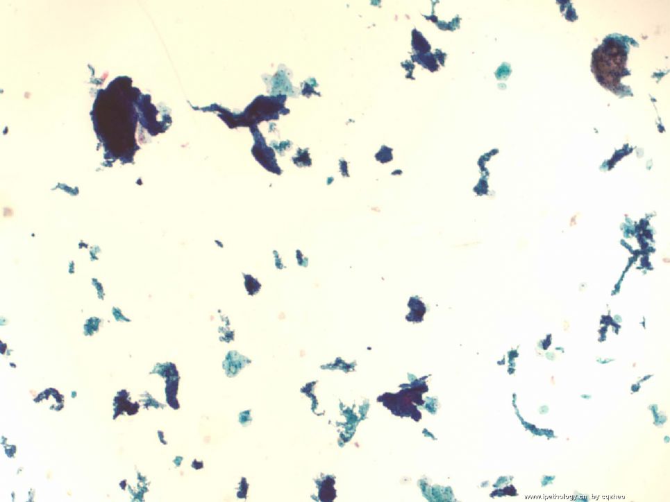
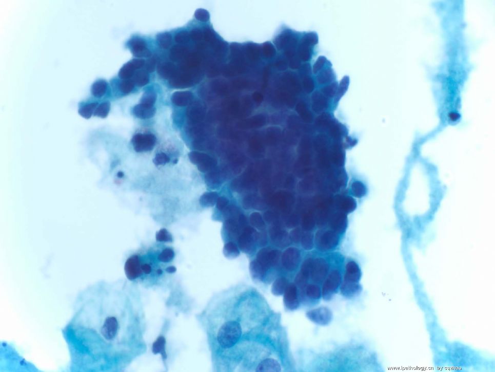
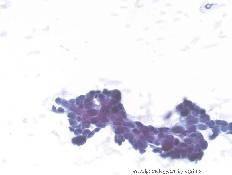
- an easy case for you-Pap test (cqz 3)
| 以下是引用月新在2008-12-30 11:03:00的发言:
译赵老师:two different interpretation above. 以上有两种不同的解释,One is likely from endometrial origin and anther one is from endocervical origin. 一种认为是子宫内膜来源,另一种认为是宫颈内膜来源。What is other pathologists' oppinion? 听一下病理医生们的意见? 我感觉两幅都没有太大的问题。第一幅象子宫内膜腺上皮细胞团,第二幅图象宫颈内膜腺上皮细胞团。如果是我的病例,但是她是一位50岁的人,建议病人一月后复查! |
Cytomorphology of Papillary Serous Adenocarcinoma
n Differential diagnosis:
1. Endometrioid carcinoma: difficult in Pap test
2. Reactive benign endocervical cells: this is the money for pathologists
3 Reactive mesothelial cells (in pelvic washing)
n clusters, flat sheets or single cells
n Cytoplasm is dense & distinct
n Multinucleation
n Nuclei chromatin vary from bland to hyperchromatic
n Nucleoli-small to prominent
Cytomorphology of Papillary Serous Adenocarcinoma (Pap)
n Malignant cells are isolated or arranged in clusters.
n Nuclei are enlarged and demonstrate variation in size.
n There is nuclear hyperchromasia with coarsely textured chromatin and prominent nucleoli.
n Cytoplasm may be scant but is often abundantly vacuolated.
n Psammoma bodies may be present.
Disease Fact Sheet about uterine serous carcinoma
1. Aggressive high grade endometrial adenocarcinoma first described in 1982.
2.
Associated with p53 mutations.
3.
Associated with endometrial polyps and commonly polypoid.
4.
Not associated with increased estrogen & endometrial hyperplasia.5. 2/3 have malignant pap smears since buds and tufts break off.
6.
25% have primary endocervical involvement.
7.
Drop metastases to the vagina is common.
8.
Transtubal spread common, creating ovarian implants and omental tumor nodules.-
It will be Christmas Eve tomorrow. I do some summaries about this Pap case
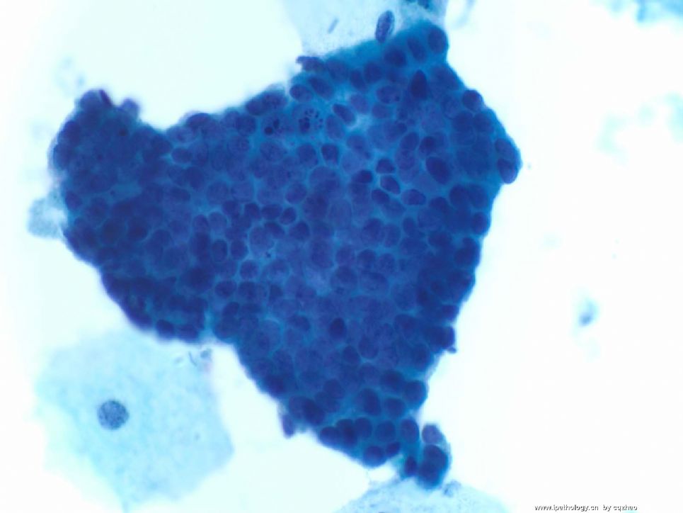
- Some of the clusters demonstrate a papillary morphology with an irregular outline.
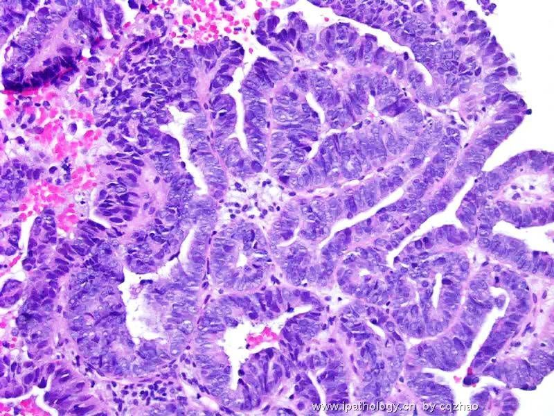
- The cell block preparation demonstrates fibrovascular cores lined by stratified malignant cells with scant cytoplasm, enlarged hyperchromatic nuclei, prominent nucleoli with scattered mitotic figures on hematoxylin and eosin staining.
Cytomorphology of the case
1. Clusters of large malignant cells with scant cytoplasm, marked variation in nuclei size, nuclear hyperchromasia with coarse chromatin, irregular nuclear membranes and prominent nucleoli.
-
本帖最后由 于 2008-12-24 12:02:00 编辑
| 以下是引用月新在2008-12-24 11:14:00的发言: 查见这样一个宫颈异形上皮细胞团,引起这么长时间的讨论,自己每发一次帖,受赵老师点评一次,感觉自己进步一次,收获很大,多年养成的小的毛病全都暴露无遗,我每次发贴都有许多暇疵,虽然干病理工作多年,工作不慎密,思维不活跃,报告不规范。这些都需要纠正。再次感谢赵老师。 |
Thank you for your good words.
You must learn Chairman Mao's words very well. You are a person with self-criticism. Kiding.
Everyone makes some mistakes, even the world-famous professors.
In the US many people always feel they are the best in the world. In fact they should learn from Chinese.
Both of your reports are very good.
Only this term 子宫内膜中分化浆液性乳头状腺癌:
Uterine serous and clear cell carcinoma are type 2 tumor. Both are high grade carcinoma. So we do not need to mention high grade serous or clear cell carcinoma. Of cause we will not say well or moderately differentiated serous or clear cell carcinoma.
Thank you two for working on this case.
Key for fig
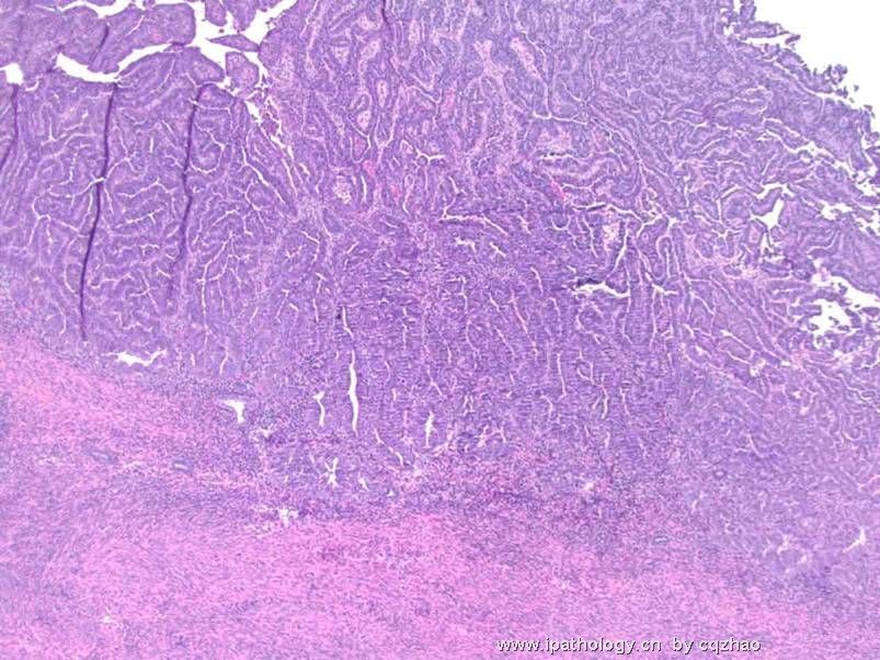
f1-2: uterus: tumor size 4x3x1.2 cm, myometrial invasion 75%.
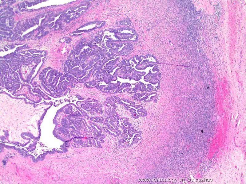
f3: R overy with focal tumor mass 4 mm, in the cortex, close to the ovary surface
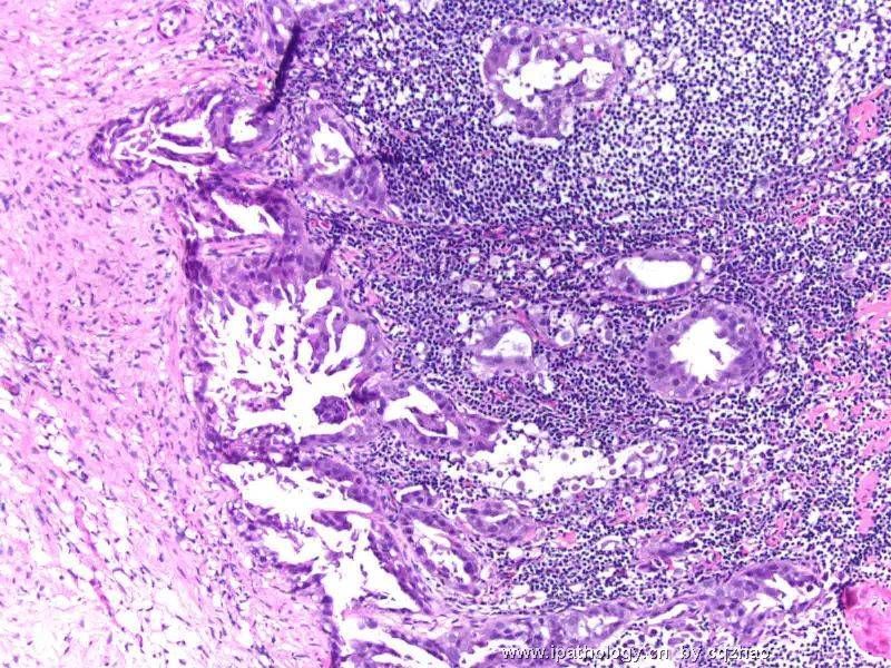
f4: omentum
f5: pelvic washing.
Lymph node is negative for malignant. No tumor is noted in the endocervix. Fallopian tubes and other ovary are normal.
How would you sign out this case with pathologic staging. I hope to see a full report. I know a lot of people here are young pathologists. You can ask your senior or professors if you are not sure. Pathologists in the US need 5 or six years of resident or fellow training (after 4 years of coolege and 4 years of medical school)and pass the difficult pathology board examination before they can have the right to sign out the cases. So it is ok to have some difficulties in pathology service if you are young and just complete medical school or jsut work few years.
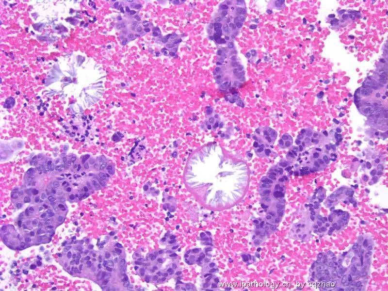
52楼图1:中间的2个管腔样结构,腔面锯齿状,看不到核,是结晶?还是上皮脱落坏死?感觉不像。还是其它病变?
Not epthelial cells. May be结晶 or other artifacts. Anyway it is not important.
图2:有乳头样结构,又有子宫内膜样癌,是混合型癌吗?可能是高级别的癌,IHC:P53,Ki67怎样?浆液性乳头状癌和子宫内膜样癌在HE上有哪些鉴别点?
It s a serous carcinoma with strong and diffuse positivity for P53. Ki67 is no use because all high grade ca can be strongly and diffusely pos. Serous ca with prominent glandular formation and without papillary features can be confused with endometrioid ca. Predominantly the nuclear features can help for the distinction. Glands in em-ca have a smooth luminal border and are lined by columinar cells with nucelar grade 1-2. EM-ca with grade 3 nuclei almost always show solid growth pattern with few glandular formation. In fact in this case you can appreciate some papillary structures even in the photo 2 of ECC specimen.
| 以下是引用月新在2008-12-19 11:05:00的发言:
|
Lesson from above case: Cell block can be very useful for diagnosis sometime.
We cannot prepare the cell blocks for all Pap test, but we may have some unexpected findings in some cases. I ask cyto lab to prepare the cell block if the Pap smears show some high grade dysplastic cells (may be carcinoma), but I am not sure for the diagnosis. In this situation you can prepare a cell block to try your luck.
