共3页/53条首页上一页123下一页尾页
回复:53 阅读:7258
B1301Uterine high grade malignant tumor with divergent differentiation (cqz3)

cqzhao 离线

帖子:5518
粉蓝豆:1071
经验:5755
注册时间:2008-09-29
加关注  |  发消息
楼主 发表于 2008-12-03 05:08|举报|关注(2)
浏览排序[ 顺序 逆序 楼主 支持 精彩 ]  快捷回复
姓    名: ××× 性别:   年龄:  
标本名称:  
简要病史:  
肉眼检查:  

Share a case of this week.

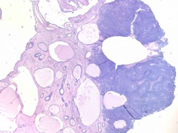
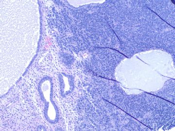
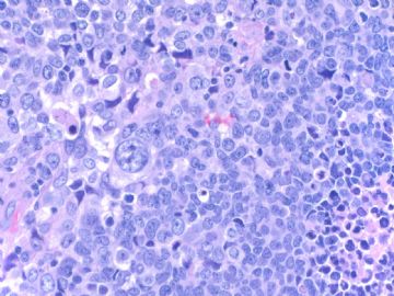
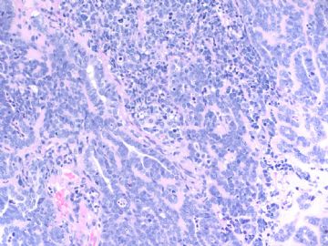
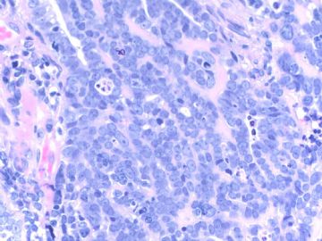
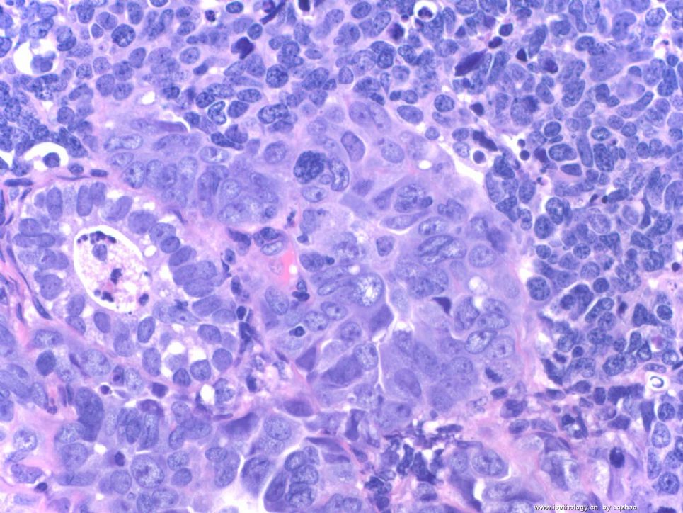
Old lady with atrophic endometrium showing tumor mass in the surface of cystic atrophic endometrium (first figure)

F1 20x

F2 100x

F3 200x

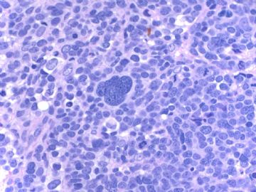
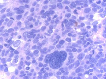
F4-5 400x

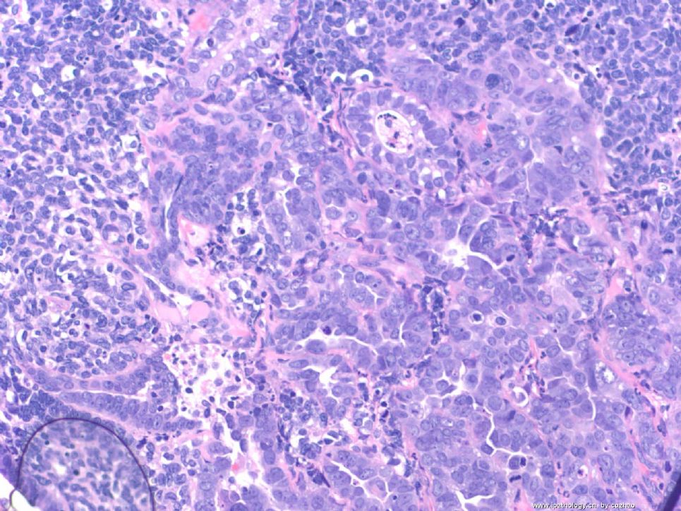
F6 200x

F7 400x

F6 and 7 showing focal glandular lesion mixed with other solid lesion.

Your dx or differential dx

  • Uterine high grade malignant tumor with divergent differentiation (cqz3)图1
    图1
  • Uterine high grade malignant tumor with divergent differentiation (cqz3)图2
    图2
  • Uterine high grade malignant tumor with divergent differentiation (cqz3)图3
    图3
  • Uterine high grade malignant tumor with divergent differentiation (cqz3)图4
    图4
  • Uterine high grade malignant tumor with divergent differentiation (cqz3)图5
    图5
  • Uterine high grade malignant tumor with divergent differentiation (cqz3)图6
    图6
  • Uterine high grade malignant tumor with divergent differentiation (cqz3)图7
    图7
标签:子宫 高级别肿瘤 异源性分化
本帖最后由 于 2009-02-25 09:51:00 编辑
0
×参考诊断
高级别肿瘤伴异源性分化

abin 离线

帖子:7416
粉蓝豆:6014
经验:11787
注册时间:2006-10-08
加关注  |  发消息
1 楼    发表于2008-12-11 23:16:00举报|引用
返回顶部 | 快捷回复
 第20楼两幅图腺癌成分与背景之间界限截然,细胞学特点也不一样,与第1楼相比,第20楼更像癌肉瘤。
0
回复
signature

华夏病理/粉蓝医疗

为基层医院病理科提供全面解决方案,

努力让人人享有便捷准确可靠的病理诊断服务。


四国迷情 离线

帖子:515
粉蓝豆:43
经验:1148
注册时间:2007-09-08
加关注  |  发消息
2 楼    发表于2008-12-06 01:09:00举报|引用
返回顶部 | 快捷回复
 支持
0
回复
signature
正在读取中……

stevenshen 离线

帖子:343
粉蓝豆:2
经验:343
注册时间:2008-06-03
加关注  |  发消息
3 楼    发表于2008-12-06 08:47:00举报|引用
返回顶部 | 快捷回复

Looks bad; guess Endometrial Intraepithelial Carcinoma (EIC)

Thanks.

0
回复

cqzhao 离线

帖子:5518
粉蓝豆:1071
经验:5755
注册时间:2008-09-29
加关注  |  发消息
4 楼    发表于2008-12-06 11:26:00举报|引用
返回顶部 | 快捷回复

 Most cases I sent here have some difficulties. Hope pathology colleaques what you will do if it is your true case. It is better to mention  your differential dx based on H&E. What IHC do you want to order based on your differential dx. In this way you can truely learn sth from studying the case.

Thanks

0
回复

cqzhao 离线

帖子:5518
粉蓝豆:1071
经验:5755
注册时间:2008-09-29
加关注  |  发消息
5 楼    发表于2008-12-06 11:31:00举报|引用
返回顶部 | 快捷回复

 EIC likes carcinoma in situ, meaning carcinoma limited within glands. EIC is considered as precursor of serous carcinoma in endometrium. Wenxing Zhang, Chinese American pathologist did a lot of research in this topic.

Cytomorphologic features of this case have no any similarity to EIC.

0
回复

cqzhao 离线

帖子:5518
粉蓝豆:1071
经验:5755
注册时间:2008-09-29
加关注  |  发消息
6 楼    发表于2008-12-06 11:33:00举报|引用
返回顶部 | 快捷回复
以下是引用wang4160在2008-12-3 17:11:00的发言:

以下是引用月新在2008-12-3 11:22:00的发言:

 局灶性的间质细胞异形性明显,有许多的核丝分裂,腺上皮感觉还好,应该考虑局灶性恶性苗勒氏管腺肉瘤。间质恶性,上皮良性。

支持!

Are you really think the glandular component in the last photos are benign?
0
回复

dujun0522 离线

帖子:406
粉蓝豆:532
经验:2696
注册时间:2008-07-11
加关注  |  发消息
7 楼    发表于2008-12-06 22:59:00举报|引用
返回顶部 | 快捷回复
 考虑癌肉瘤。
0
回复

njwbhuang 离线

帖子:712
粉蓝豆:2265
经验:1404
注册时间:2007-11-23
加关注  |  发消息
8 楼    发表于2008-12-07 20:31:00举报|引用
返回顶部 | 快捷回复
以下是引用cqzhao在2008-12-6 11:26:00的发言:

 Most cases I sent here have some difficulties. Hope pathology colleaques what you will do if it is your true case. It is better to mention  your differential dx based on H&E. What IHC do you want to order based on your differential dx. In this way you can truely learn sth from studying the case.

Thanks

译文:我提供的大多数病例都是有一些难度。希望病理同仁应该做到这样:即如果该病例是我的,我该怎么做。根据HE切片提出你的鉴别诊断,然后选择哪些免疫标记,只有这样你才能从该病例中学到东西。


非常赞同赵老师的意见。

0
回复

stevenshen 离线

帖子:343
粉蓝豆:2
经验:343
注册时间:2008-06-03
加关注  |  发消息
9 楼    发表于2008-12-08 22:26:00举报|引用
返回顶部 | 快捷回复

Great advice! But I like and enjoy taking a shot at any case without fear of embarrassment, particularly with a few photos. I would encourage the readers (younger pathologists) to do the same. This is why this forum has a lot of fun!

0
回复

天山望月 离线

帖子:4902
粉蓝豆:83
经验:5092
注册时间:2007-02-03
加关注  |  发消息
10 楼    发表于2008-12-08 22:52:00举报|引用
返回顶部 | 快捷回复

 非常赞同Dr.zhao 和Dr.shen 的建议!

有时没有写下诊断,是想听听大家的意见,不想先入为主,误导。讨论中可以写上自己的观点。仅为个人观点

0
回复
signature
广州金域病理

cqzhao 离线

帖子:5518
粉蓝豆:1071
经验:5755
注册时间:2008-09-29
加关注  |  发消息
11 楼    发表于2008-12-09 02:38:00举报|引用
返回顶部 | 快捷回复
 Agree with  above. You can say what you want to say and do not have any responsbility. No person will remember who is right and who is wrong. Just imagin you can fly in the sky. Ha, ha
0
回复

陈隆文博士 离线

帖子:180
粉蓝豆:4
经验:182
注册时间:2008-08-27
加关注  |  发消息
12 楼    发表于2008-12-09 06:23:00举报|引用
返回顶部 | 快捷回复
本帖最后由 于 2008-12-11 23:35:00 编辑

All the cases posted by Dr. Zhao are pretty interesting. This case is no exception. Basically, we all agree that it is a high-grade malignancy and it mostly likely involve an endometrial polyp (fist figure).  There are mainly several possibilities: 1) a high nuclear grade carcinoma (such as serous carcinoma or undifferentiated carcinoma); 2) Carcinosarcoma (癌肉瘤); 3) Adenosracoma (腺肉瘤) with sarcomatous overgrowth. I don't think that this case fits well with adenosarcoma, since I favor there is at least some adenocarcinomatous components. I did not realize that even expert GYN pathologists cannot always agree on the diagnosis of Cracinosarcoma until I came to Cleveland Clinic and learned GYN path from Dr. Bill Hart (one of the top GYN pathologists in the USA). He has a very strigent criteria for Carcinosarcoma, which should have a perfect bi-phasic pattern of malignant glands and stroma. I doubt that he will call this thing carcinosarcoma.

 For all practical purpose, we would NOT do a lot immunostains in  biopsy cases in our practice, since the clinical management does not matter, if you call high-grade carcinoma vs. carcinosarcoma or even adenosarcoma with sarcomatous overgrowth, it will be total hystrectomy with lymph node staging. So, we usually further classify a tumor on the bigger resection specimen. Not uncommon, you thought it was high-grade carcinoma on the biopsy, but when the whole uterus came out, it clearly showed carcinosarcoma. Is this a biopsy (curretings) or hysterectomy? Thanks!

abin译:

Dr. Zhao提供的病例都非常有趣,这一例也不例外。我们基本上都一致认为它是高级别恶性肿瘤,并且它可能累犯子宫内膜息肉(第1图)。有几种可能性:1)高核级别的癌(如浆液性癌或未分化癌);2)癌肉瘤;3)腺肉瘤伴肉瘤样过度生长。我认为不太像腺癌,我倾向于至少有部分腺癌样成分。在我来到Cleveland Clinic并向Dr. Bill Hart(美国顶级妇科病理专家之一)学习妇科病理之前,不知道即使妇科病理专家对癌肉瘤的诊断也不一致。他对癌肉瘤有非常严格的诊断标准,即:恶性腺体和间质必须形成非常完美的双相性形态。

从实用角度看,我们不会对活检标本做许多免疫染色,因为临床处理差别不大,如果称为高级别癌vs癌肉瘤vs腺癌伴肉瘤样过度生长,都是全子宫切除加淋巴结分期。因此,我们通常在较大标本上作进一步分类。不少情况下,活检认为是高级别癌,而全子宫标本变成明显的癌肉瘤。这是活检(诊刮)还是子宫切除标本?谢谢!

0
回复

cqzhao 离线

帖子:5518
粉蓝豆:1071
经验:5755
注册时间:2008-09-29
加关注  |  发消息
13 楼    发表于2008-12-09 09:57:00举报|引用
返回顶部 | 快捷回复
 Thank Dr. Chen's excellent analysis. This is hysterectomy specimen with several tumor nodules as fig. Pt had biopsy.
0
回复

alading1999 离线

帖子:63
粉蓝豆:31
经验:562
注册时间:2009-01-17
加关注  |  发消息
14 楼    发表于2009-03-11 12:28:00举报|引用
返回顶部 | 快捷回复
 很精彩!!谢谢各位老师。。
0
回复

fuying 离线

帖子:362
粉蓝豆:2
经验:390
注册时间:2008-04-10
加关注  |  发消息
15 楼    发表于2008-12-03 09:51:00举报|引用
返回顶部 | 快捷回复
本帖最后由 于 2008-12-03 14:56:00 编辑  少见病例,病灶局限,间质细胞和内膜腺上皮细胞均有不同程度的异型性。
0
回复

月新 离线

帖子:528
粉蓝豆:1313
经验:547
注册时间:2006-10-05
加关注  |  发消息
16 楼    发表于2008-12-09 22:23:00举报|引用
返回顶部 | 快捷回复
 感谢赵老师继续,
0
回复

wang4160 离线

帖子:2256
粉蓝豆:16
经验:2617
注册时间:2007-01-31
加关注  |  发消息
17 楼    发表于2008-12-03 17:11:00举报|引用
返回顶部 | 快捷回复
以下是引用月新在2008-12-3 11:22:00的发言:

 局灶性的间质细胞异形性明显,有许多的核丝分裂,腺上皮感觉还好,应该考虑局灶性恶性苗勒氏管腺肉瘤。间质恶性,上皮良性。

支持!
0
回复

njwbhuang 离线

帖子:712
粉蓝豆:2265
经验:1404
注册时间:2007-11-23
加关注  |  发消息
18 楼    发表于2008-12-03 20:25:00举报|引用
返回顶部 | 快捷回复
 病变位于萎缩性子宫内膜的一侧,低倍镜下可见瘤细胞呈片状或实性巢状排列,中倍和高倍可见瘤细胞胞质较少,核卵圆形或圆形,染色质粉尘状,可见核仁和核分裂,其间见有一个肿瘤性多核巨细胞。部分肿瘤细胞呈腺样排列。根据这些图片,主要考虑为子宫内膜未分化癌,由于肿瘤位于子宫内膜间质中,子宫内膜间质肉瘤需要排除。组织学图像非常像肺的小细胞癌,子宫内膜原发性或转移性小细胞癌要考虑。
0
回复

abin 离线

帖子:7416
粉蓝豆:6014
经验:11787
注册时间:2006-10-08
加关注  |  发消息
19 楼    发表于2008-12-11 12:30:00举报|引用
返回顶部 | 快捷回复

 很有趣的病例!Dr.cqzhao的宝贝真多,学不完哈!

图像显示老年女性萎缩子宫内膜背景上出现的恶性形态成分。最后二图可以辨认腺样分化,并且与实性成分移行过渡。因此考虑:低分化or未分化or去分化腺癌,是否存在肉瘤成分可能需要免疫组化帮助,仅用CK和Vimentin也许足够了。我感觉没有肉瘤成分。

它不是EIC。在郑文新教授的启发下,我也做一些UPSC及其前驱病变的研究工作。

0
回复
signature

华夏病理/粉蓝医疗

为基层医院病理科提供全面解决方案,

努力让人人享有便捷准确可靠的病理诊断服务。


cqzhao 离线

帖子:5518
粉蓝豆:1071
经验:5755
注册时间:2008-09-29
加关注  |  发消息
20 楼    发表于2008-12-11 04:26:00举报|引用
返回顶部 | 快捷回复
本帖最后由 于 2008-12-11 04:29:00 编辑

 took two previous endometrial biopsy photos

200x

400x


名称:图1
描述:图1

名称:图2
描述:图2
0
回复
回复:53 阅读:7258
共3页/53条首页上一页123下一页尾页
【免责声明】讨论内容仅作学术交流之用,不作为诊疗依据,由此而引起的法律问题作者及本站不承担任何责任。
快速回复
进入高级回复
您最多可输入10000个汉字,按 "Ctrl" + "Enter" 直接发送
搜索回复/乘电梯 ×
按内容
按会员
乘电梯
合作伙伴
友情链接