
图1")
图2")
图3")
图4")
图5")
图6")
图7")


| 图片: | |
|---|---|
| 名称: | |
| 描述: | |
- B1301Uterine high grade malignant tumor with divergent differentiation (cqz3)
图1") 图1
图1图2") 图2
图2图3") 图3
图3图4") 图4
图4图5") 图5
图5图6") 图6
图6图7") 图7
图7
| 姓 名: | ××× | 性别: | 年龄: | ||
| 标本名称: | |||||
| 简要病史: | |||||
| 肉眼检查: | |||||
Share a case of this week.
Old lady with atrophic endometrium showing tumor mass in the surface of cystic atrophic endometrium (first figure)
F1 20x
F2 100x
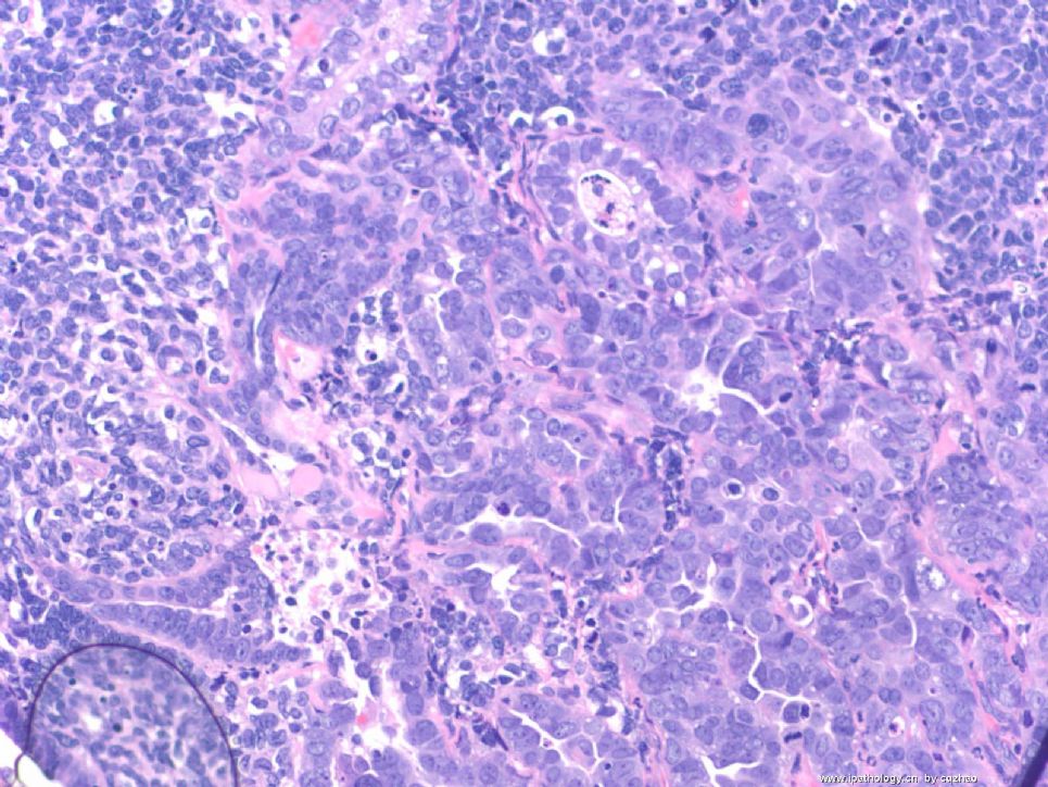
F3 200x
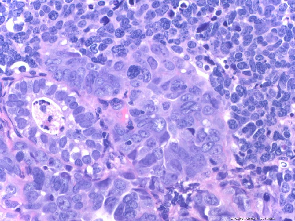
F4-5 400x
F6 200x
F7 400x
F6 and 7 showing focal glandular lesion mixed with other solid lesion.
Your dx or differential dx
标签:子宫 高级别肿瘤 异源性分化
-
本帖最后由 于 2009-02-25 09:51:00 编辑
相关帖子
- • 来一例简单罕见的(有诊断)
- • 是肉瘤吗?
- • 子宫内膜,复杂性增生?癌?(有大体结果了)
- • 子宫肿物(透明细胞平滑肌瘤?)
- • 子宫肌壁间肿物
- • 子宫腔内占位。
- • 子宫肿瘤
- • 子宫肌层浸润性癌
- • 8 个子宫上皮肿瘤病例-扫描图片
- • 子宫平滑肌肿瘤?间质肉瘤?
×参考诊断
高级别肿瘤伴异源性分化
-
stevenshen 离线
- 帖子:343
- 粉蓝豆:2
- 经验:343
- 注册时间:2008-06-03
- 加关注 | 发消息
Most cases I sent here have some difficulties. Hope pathology colleaques what you will do if it is your true case. It is better to mention your differential dx based on H&E. What IHC do you want to order based on your differential dx. In this way you can truely learn sth from studying the case.
Thanks
EIC likes carcinoma in situ, meaning carcinoma limited within glands. EIC is considered as precursor of serous carcinoma in endometrium. Wenxing Zhang, Chinese American pathologist did a lot of research in this topic.
Cytomorphologic features of this case have no any similarity to EIC.
| 以下是引用cqzhao在2008-12-6 11:26:00的发言:
Most cases I sent here have some difficulties. Hope pathology colleaques what you will do if it is your true case. It is better to mention your differential dx based on H&E. What IHC do you want to order based on your differential dx. In this way you can truely learn sth from studying the case. Thanks |
译文:我提供的大多数病例都是有一些难度。希望病理同仁应该做到这样:即如果该病例是我的,我该怎么做。根据HE切片提出你的鉴别诊断,然后选择哪些免疫标记,只有这样你才能从该病例中学到东西。
非常赞同赵老师的意见。
-
stevenshen 离线
- 帖子:343
- 粉蓝豆:2
- 经验:343
- 注册时间:2008-06-03
- 加关注 | 发消息
-
本帖最后由 于 2008-12-11 23:35:00 编辑
All the cases posted by Dr. Zhao are pretty interesting. This case is no exception. Basically, we all agree that it is a high-grade malignancy and it mostly likely involve an endometrial polyp (fist figure). There are mainly several possibilities: 1) a high nuclear grade carcinoma (such as serous carcinoma or undifferentiated carcinoma); 2) Carcinosarcoma (癌肉瘤); 3) Adenosracoma (腺肉瘤) with sarcomatous overgrowth. I don't think that this case fits well with adenosarcoma, since I favor there is at least some adenocarcinomatous components. I did not realize that even expert GYN pathologists cannot always agree on the diagnosis of Cracinosarcoma until I came to Cleveland Clinic and learned GYN path from Dr. Bill Hart (one of the top GYN pathologists in the USA). He has a very strigent criteria for Carcinosarcoma, which should have a perfect bi-phasic pattern of malignant glands and stroma. I doubt that he will call this thing carcinosarcoma.
For all practical purpose, we would NOT do a lot immunostains in biopsy cases in our practice, since the clinical management does not matter, if you call high-grade carcinoma vs. carcinosarcoma or even adenosarcoma with sarcomatous overgrowth, it will be total hystrectomy with lymph node staging. So, we usually further classify a tumor on the bigger resection specimen. Not uncommon, you thought it was high-grade carcinoma on the biopsy, but when the whole uterus came out, it clearly showed carcinosarcoma. Is this a biopsy (curretings) or hysterectomy? Thanks!
abin译:
Dr. Zhao提供的病例都非常有趣,这一例也不例外。我们基本上都一致认为它是高级别恶性肿瘤,并且它可能累犯子宫内膜息肉(第1图)。有几种可能性:1)高核级别的癌(如浆液性癌或未分化癌);2)癌肉瘤;3)腺肉瘤伴肉瘤样过度生长。我认为不太像腺癌,我倾向于至少有部分腺癌样成分。在我来到Cleveland Clinic并向Dr. Bill Hart(美国顶级妇科病理专家之一)学习妇科病理之前,不知道即使妇科病理专家对癌肉瘤的诊断也不一致。他对癌肉瘤有非常严格的诊断标准,即:恶性腺体和间质必须形成非常完美的双相性形态。
从实用角度看,我们不会对活检标本做许多免疫染色,因为临床处理差别不大,如果称为高级别癌vs癌肉瘤vs腺癌伴肉瘤样过度生长,都是全子宫切除加淋巴结分期。因此,我们通常在较大标本上作进一步分类。不少情况下,活检认为是高级别癌,而全子宫标本变成明显的癌肉瘤。这是活检(诊刮)还是子宫切除标本?谢谢!


