| 图片: | |
|---|---|
| 名称: | |
| 描述: | |
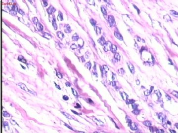
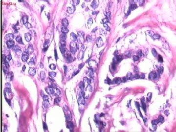
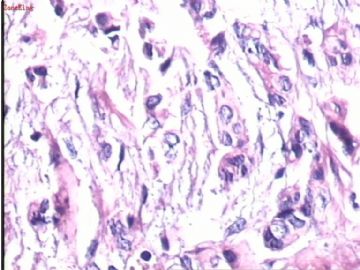
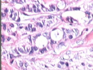
- B1732女,53岁,乳腺肿块--已上传免疫组化结果。
| 姓 名: | ××× | 性别: | 女 | 年龄: | 53岁 |
| 标本名称: | 乳腺肿块 | ||||
| 简要病史: | 发现右乳腺外上象限肿块半年余 | ||||
| 肉眼检查: | 灰白黄色组织4x3.5x3.5cm,切开,切面有一肿块,直径2.5cm,无包膜,灰白淡红色,疤痕样浸润性生长,质硬,挤压无液体流出。 | ||||
-
本帖最后由 于 2008-12-01 19:30:00 编辑

- 广州金域病理
相关帖子
-
本帖最后由 于 2008-12-10 22:54:00 编辑
Thank 天山望月 for the good case. It is invasive ductal carcinoma based on your IHC result. Please always do IHC if you are not sure the nature of the tumors. Otherwise you will be fooled sometimes. I learn this from my mistakes.
(abin译:谢谢天山望月提供的好病例。根据你的免疫组化结果,这例是浸润性导管癌。如果不确定肿瘤性质,请做免疫组化。否则有时会中招。我是从自己的失误中得到的教训。)
-
本帖最后由 于 2008-12-10 22:51:00 编辑
| 以下是引用abin在2008-11-22 18:29:00的发言:
既有浸润性小叶癌特点,又有浸润性导管癌特点 免疫组化确定是否存在浸润性小叶癌成分;如果有,这例是混合性浸润性导管癌和浸润性小叶癌。如果没有免疫组化,考虑具有浸润性小叶癌特点的浸润性乳腺癌 |
Agree with Abin.
The morphology is consistent with invasive lobular ca. However some high power photos show clearly glandular formation, looking like invasive ductal ca. I will not consider tubular ca in this case.
We will always do E-cadherine and p120 for this kind of cases to make sure it is IDC, ILC or mixed type.
(abin译:
同意。
形态学符合浸润性小叶癌。然而一些高倍图示明显的腺样结构,像浸润性导管癌。本例我不考虑小管癌。我们常用C-Ca和p120来明确是IDC,ILC或二者混合。)
-
lfl001200546 离线
- 帖子:2808
- 粉蓝豆:40
- 经验:2808
- 注册时间:2007-02-14
- 加关注 | 发消息

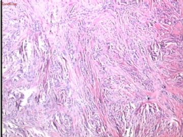
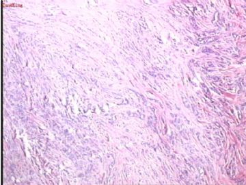
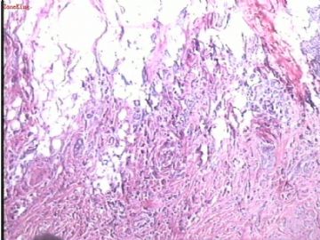
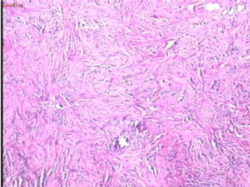
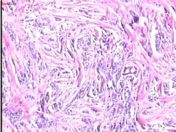
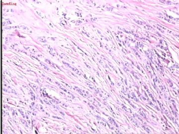
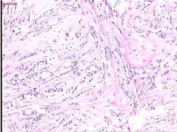
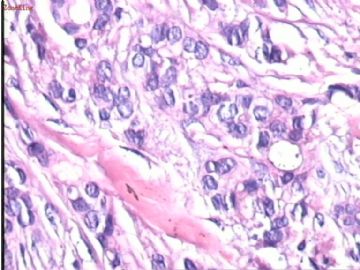
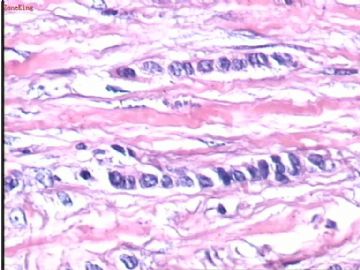
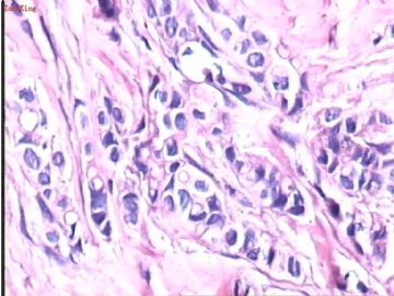
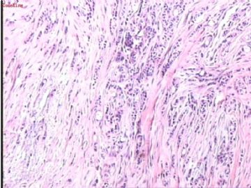
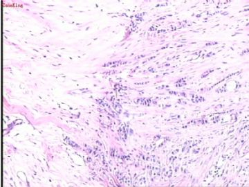
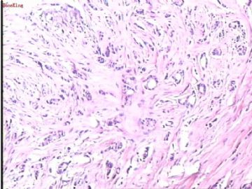
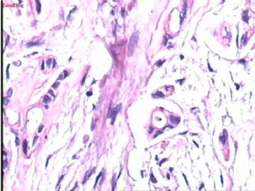
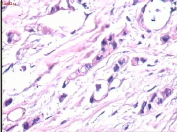


 也可能是间质硬化明显的浸润性导管癌,过去所谓的硬癌。


