| 图片: | |
|---|---|
| 名称: | |
| 描述: | |
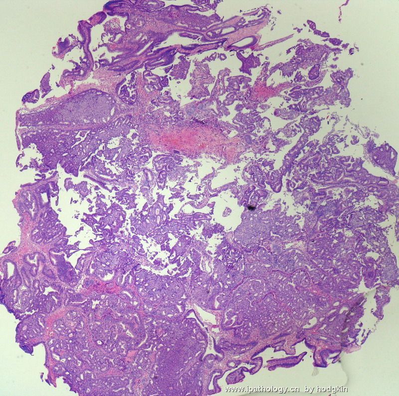
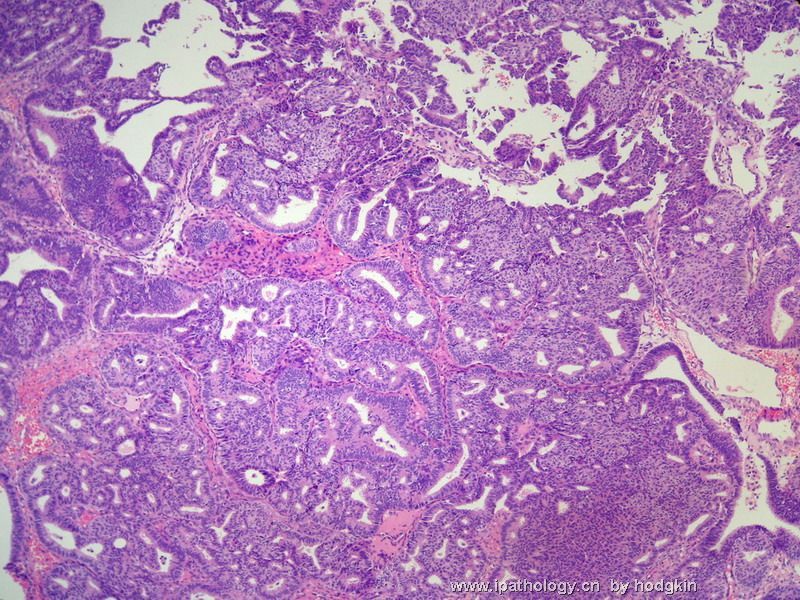
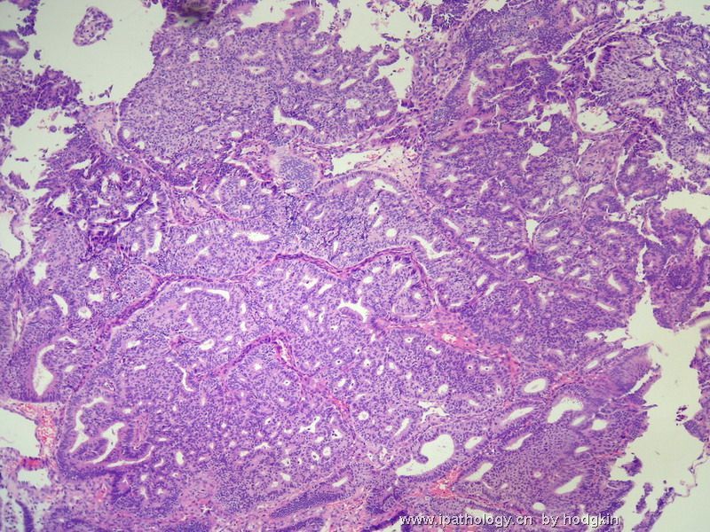
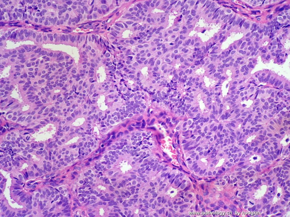
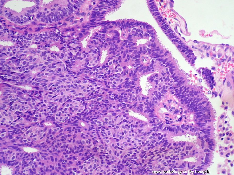
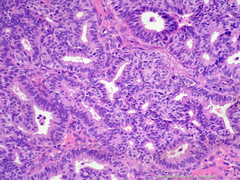
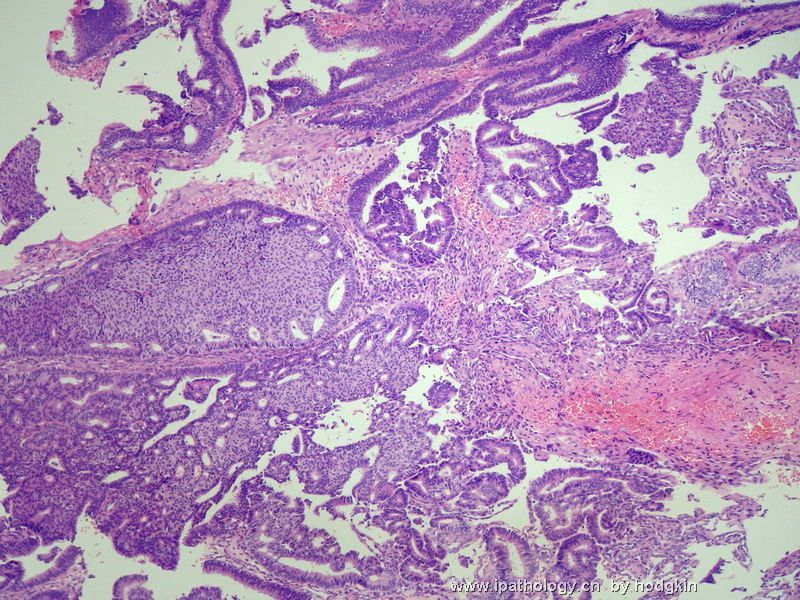
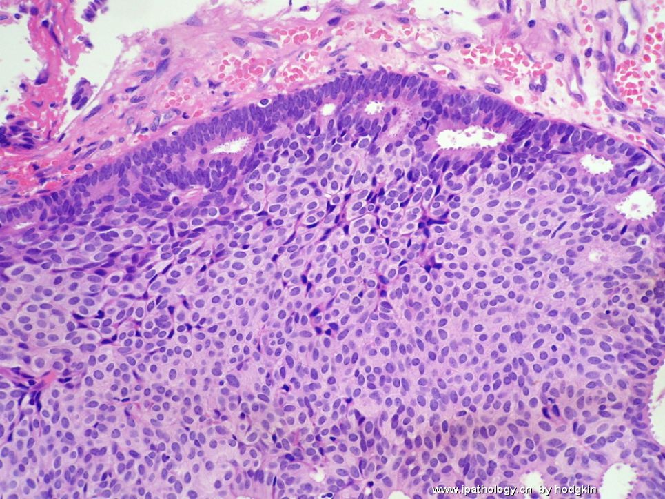
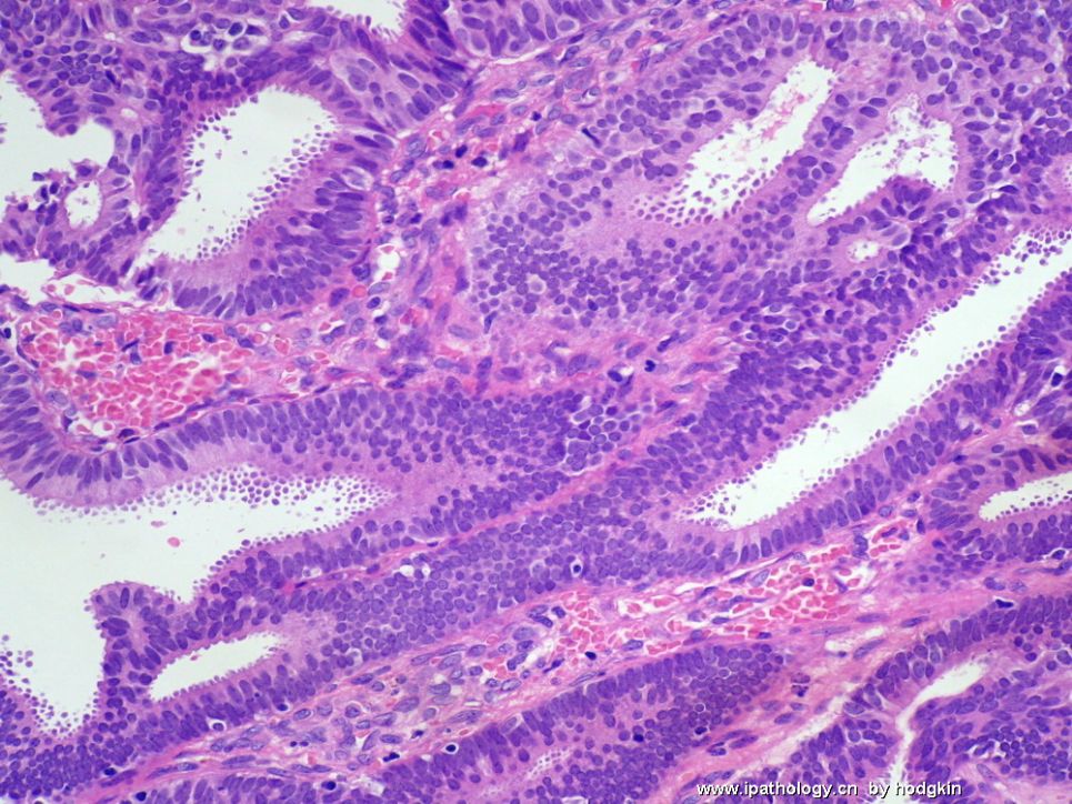
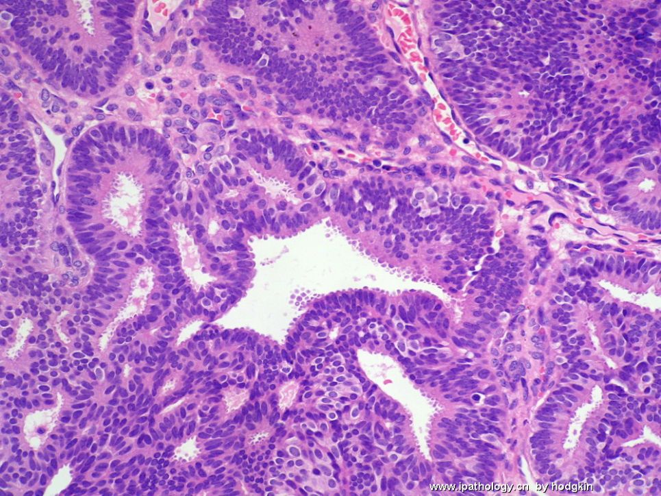
- 57岁女性,乳腺肿物。
-
baixuefeng 离线
- 帖子:73
- 粉蓝豆:422
- 经验:411
- 注册时间:2006-10-21
- 加关注 | 发消息
-
stevenshen 离线
- 帖子:343
- 粉蓝豆:2
- 经验:343
- 注册时间:2008-06-03
- 加关注 | 发消息
-
本帖最后由 于 2008-12-08 18:41:00 编辑
I have a slight different opinion regarding UDH-ADH-DCIS, I don't consider them entirely continuous, UDH share morphologic similarities with ADH, but I would consider UDH completely different from ADH/low grade DCIS. ADH/DCIS are clonal neoplastic process, but UDH is a proliferative non-neoplastic process. Thanks.
abin译:
UDH-ADH-DCIS并不是完全连续的过程。UDH与ADH形态相似,但我认为UDH与ADH/低级别DCIS完全不同。ADH/DCIS是克隆性肿瘤性病变,而UDH是增生性非肿瘤性病变。
-
Anyway this is a interesting case. The key for this case is ADH or DCIS. It is a continueous process from UDH-ADH-DCIS. We arbitrarily divide them into different categories based the standards. Of cause there are some subjective factors. It is not unusual that there are poor agreement between experter pathologists for some borderline cases.
It is an interesting case. There are so many people joining the discussion with variable oppinion, UDH, ADH, DCIS, invasive ca. Some of you changed your diagnosis after reading the case again, such as Dr. 天山望月
The photos demonstrate extensive ductal hyperplasia and columnar cell change. Some ducts show florid ductal hyperplasia with a substantial solid growth and with fenestrations distributed at the periphery. Overall proliferation tends more cellular and complex. Foci of florid ductal hyperplasia are more likely to fill the entire duct lumen in a solid or fenestrated fashion. Nuclei are overlapping and streaming. These are classic characteristic features of florid ductal hyperplasia. Focal ductal proliferation shows monomorphic cells with feature suggestive of (or consistent with) atypical ductal hyperplasia. I do not see any feature of DCIS in this case.
ER stains ductal epithelial cells and SMA or CK5/6 stain myoepithelial cells. Florid ductal hyperplasia with solid pattern means proliferative ductal cells almost fill in the entire ducts and without myoepithelial cells. Of cause ER stain is diffusely positive and myoepithelial markers are negative within the ducts. I repeated many times that IHC will not help you for differental dx of udh, adh dcis. I know your guys still do not believe it.
Is this an invasive ca? It does not look like inv ca based on the growth pattern and cytologic features in H&E. I can not ppreciate the SMA stain in frozen section and also do not thust the stain in frozen. You want to diagnose invasion in this case you must show the good myoepithelial stains in perminent sections.
My diagnosis again (I mentioned before):
Focal ADH
florid ductal hyperplasia
Columnar cell change
I do not know how many people I can convince after the discussion.

