| 图片: | |
|---|---|
| 名称: | |
| 描述: | |
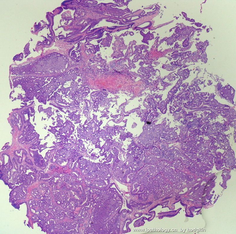
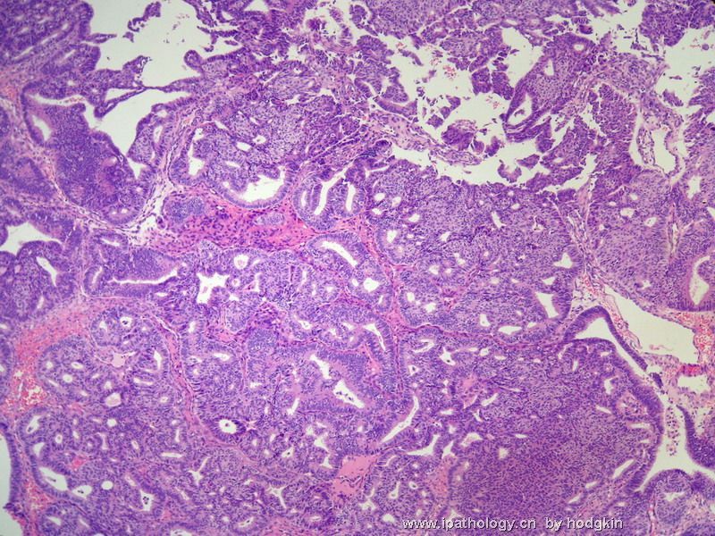
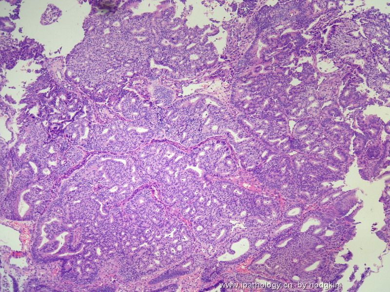
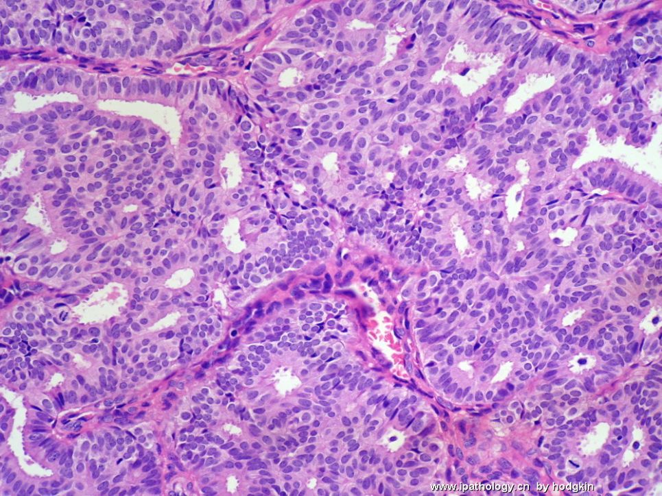
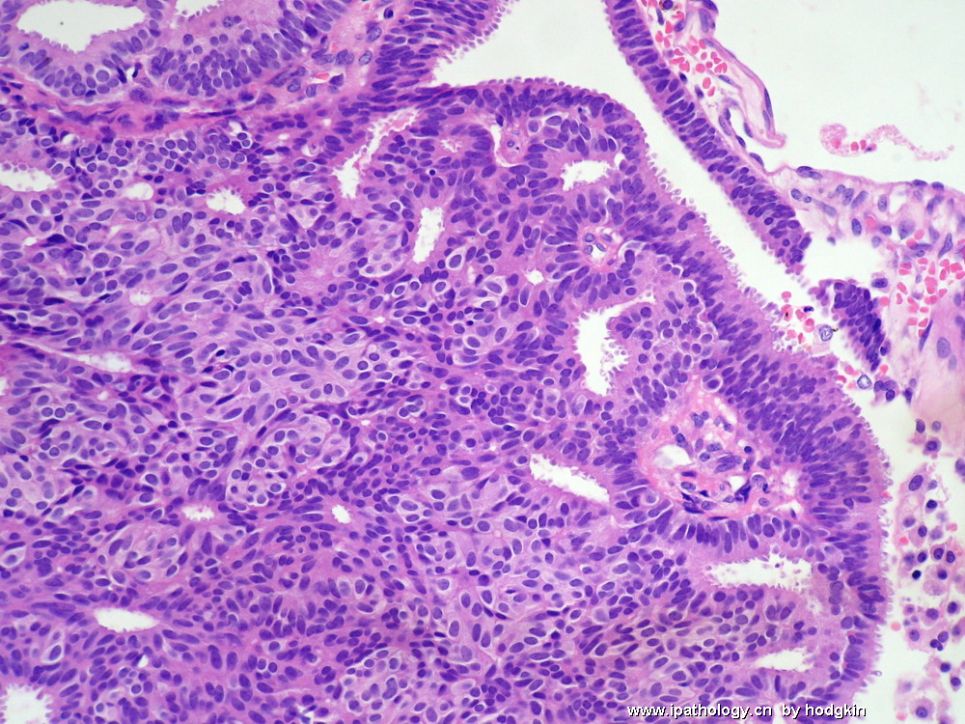
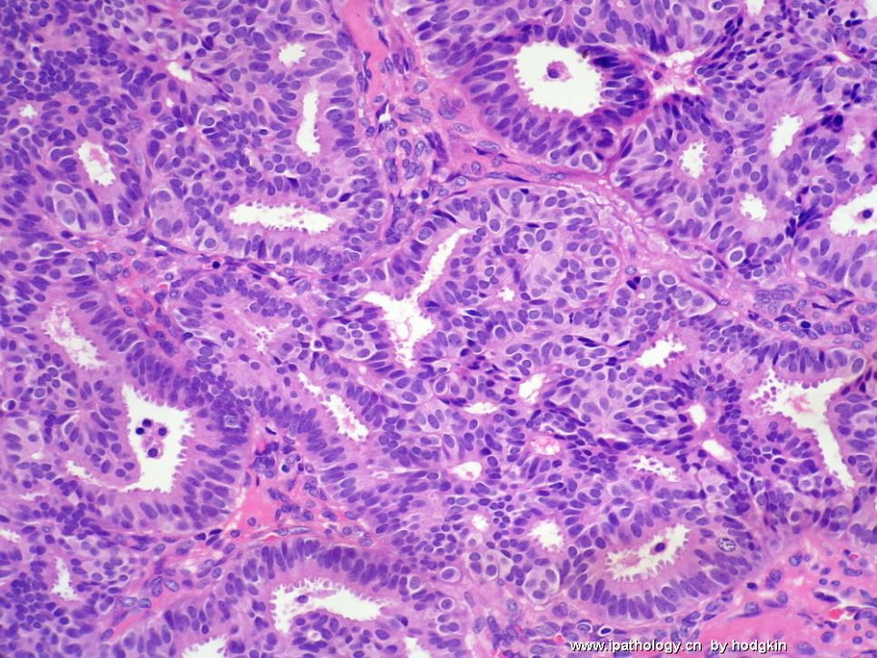
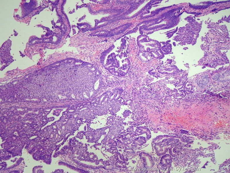
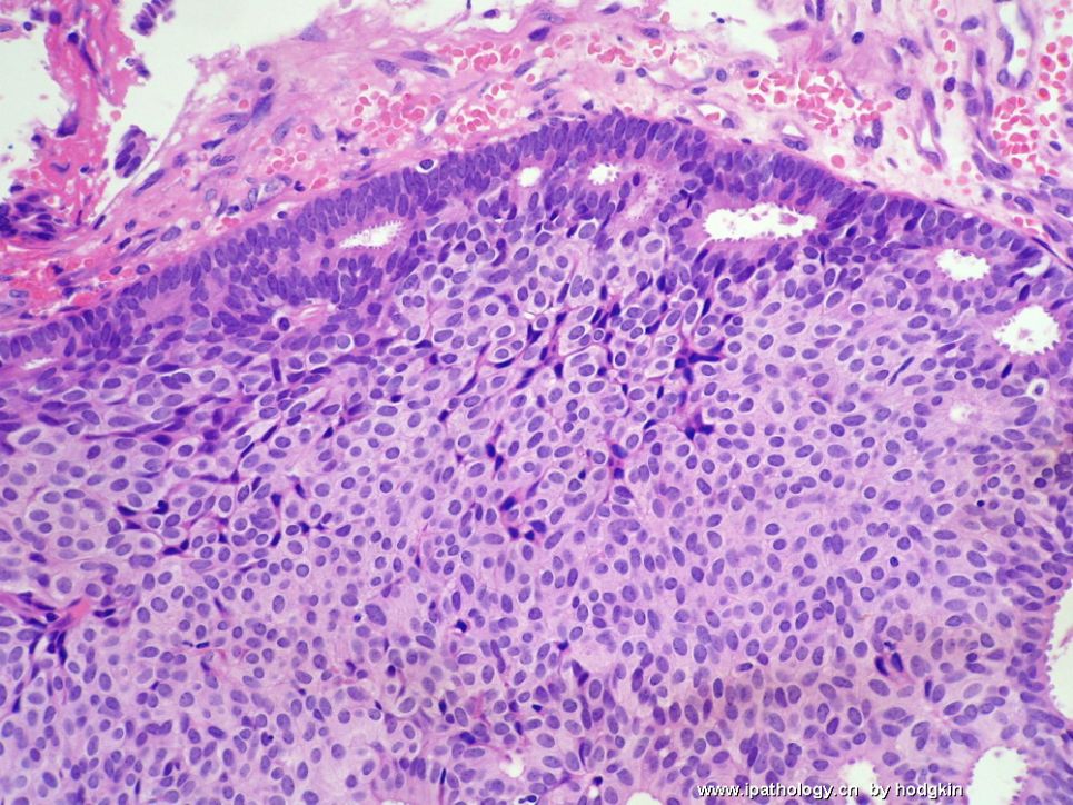
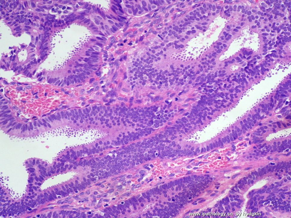
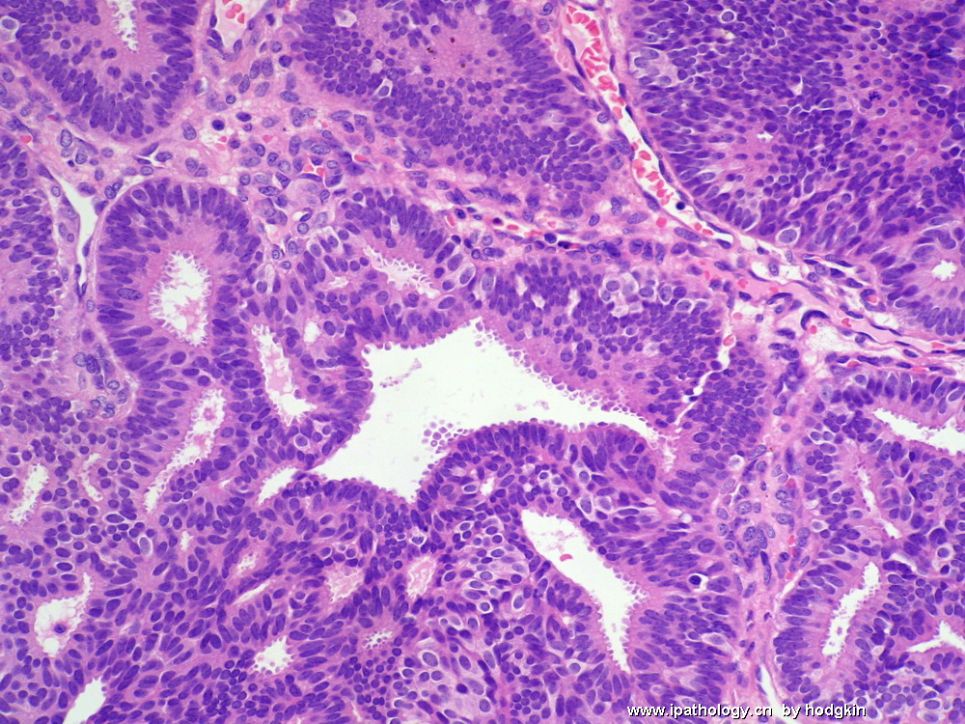
- 57岁女性,乳腺肿物。
-
Anyway this is a interesting case. The key for this case is ADH or DCIS. It is a continueous process from UDH-ADH-DCIS. We arbitrarily divide them into different categories based the standards. Of cause there are some subjective factors. It is not unusual that there are poor agreement between experter pathologists for some borderline cases.
-
stevenshen 离线
- 帖子:343
- 粉蓝豆:2
- 经验:343
- 注册时间:2008-06-03
- 加关注 | 发消息
-
本帖最后由 于 2008-12-08 18:41:00 编辑
I have a slight different opinion regarding UDH-ADH-DCIS, I don't consider them entirely continuous, UDH share morphologic similarities with ADH, but I would consider UDH completely different from ADH/low grade DCIS. ADH/DCIS are clonal neoplastic process, but UDH is a proliferative non-neoplastic process. Thanks.
abin译:
UDH-ADH-DCIS并不是完全连续的过程。UDH与ADH形态相似,但我认为UDH与ADH/低级别DCIS完全不同。ADH/DCIS是克隆性肿瘤性病变,而UDH是增生性非肿瘤性病变。
-
baixuefeng 离线
- 帖子:73
- 粉蓝豆:422
- 经验:411
- 注册时间:2006-10-21
- 加关注 | 发消息
I am surprised to see so many people are interested in this case.
Again, ADH and DCIS can be a continous lesions.
ADH is an epithelial proliferation confined to the mammary ductal-lobula system and composed in part of a neoplastic cell population similar to that seen in low-grade DCIS. However, the involved spaces also contain a population of cells more characteristic of UDH or residual normal epithelial. A diagnosis of ADH should be only to lesions in which the diagnosis of low grade DCIS is seriosly considered, but in which the features are not sufficientlly developed for a definite dx of DCIS. Some authorities also include in the definition of ADH lesions that have all of the cytologic architectural features of low grade DCIS that are limited in extent. So there are two criteria: quantitative and qualitative. There are no universally accepted size cutoff for the distinction of ADH from low grade DCIS. Tavassoli and Norris chose <2mm as ADH (Tavassoli FA, Norris HJ. Cancer 1990;65:518-529). Dr. Page originally proposed that all features of low grade DCIS be presnt in at least two separate spaces before a dx of DCIS is rendered and that anything less be considered as ADH (Page DL et al. Cancer 1985;55:2698-2708). More recently Page suggested a cutoff of 2 to 3 mm for the distiction (Jensen R, Page DL The breast. Edinburgh: Churchil Livigstone. 1998:65-89). Dr. Paul Peter Rosen in his "Rosen's Breast Pathology" (Lippincott williams and Wilkins. Second edition 2001;213) ( now there is the third edition already, best book I think) mentioned that the foregoing quantitative criteria are arbitrary and lack biologic validation. There are no a prior reason for choosing two ductal cross sections or 2 mm as critical decision points in relation to risk. No published scientific studies have compared the clinical significance of different quantitative criteria.
Now you know why it is difficult to distinguish the two lesions. We have 15 breast.gyncologic pathologists in our department. We have disagreement for some cases among pathologists in our dept. In my clinical pratice, I make the dx based on primarily the qualitative features. However the size of the lesion is also taken into the consideration. I try to overdiagnosis. Moreover, I consider the the type s of specimens. I may call ADH more for some borderline cases in the core biopsy specimens. In our hospital the patients will have the same procedures----excisional biopsy for ADH or focal low grade DCIS in core biopsy. So you should make your judgment based on the criteria (no universal one), patients' situation, communication with surgens, your experience. Should avoid overlabeling the patient as having cancer.
Finely the size criteria are used only to the distinction of ADH from low grade DCIS. Intermediate (nuclear grade 2) or high grade (nuclear grade 3) DCIS should be diagnosed as DCIS, even if present in a single space, or less than 2 mm.
I feel that I am writing a paper discussion. Hope it can have some help for you.
翻译75楼内容:
很惊奇这么多人对本例有兴趣。
重复一下,ADH和DCIS可能是连续的病变。
ADH是局限于乳腺导管-小叶系统(即TDLU,译者注)的导管上皮增生性病变,部分细胞由相似于低级别DCIS的肿瘤细胞群组成。然而,它累及的范围之内也包含类似于UDH中较为特征性的导管上皮或类似于残留的正常导管上皮。 诊断ADH仅用于以下这一情形:非常担心存在低级别DCIS,但是其特征不足以明确诊断DCIS。一些作者也将具有低级别DCIS的细胞学形态和组织结构但是范围有限定义为ADH病变。因此有两个标准:质和量。ADH与低级别DCIS之间的大小界限没有公认的结构。Tavassoli and Norris选择<2mm作为ADH (Tavassoli FA, Norris HJ. Cancer 1990;65:518-529)。Dr. Page最初提出至少两个分隔部位存在所有低级别DCIS特征,但其它方面不足以诊断者,为ADH(Page DL et al. Cancer 1985;55:2698-2708)。最近Page建议2 to 3 mm作为二者的区分。(Jensen R, Page DL The breast. Edinburgh: Churchil Livigstone. 1998:65-89)。 Dr. Paul Peter Rosen所著的《Rosen's Breast Pathology》(Lippincott williams and Wilkins. Second edition 2001;213,现在已有第三版,我认为是最好的乳腺病理专著)提到以前的量化标准都是主观的,没有明确的生物学意义。也没有明显的理由认为选择两个导管横切面或2mm作为与风险有关的严格定义。目前还没有科学研究报道比较不同量化标准的临床意义。
现在你明白为什么很难区分这两种病变。我们科有15位乳腺/妇科病理学家,我们科的病理学家对一些病变认识也不一致。在我的临床实践中,我根据质的特征作出诊断。然而病变大小也要考虑。我尽量过诊断。而且,我也考虑标本的类型。在粗针穿刺活检标本中,我可能更多使用ADH。在我们医院,粗针穿刺活检诊断的ADH或局灶性低级别DCIS,其临床处理相同:均进行切除活检。因此你可以在标准(没有公认的标准之一)、患者情形、与外科医生的交流以及自己的经验之间做出判断。应当避免过诊断为恶性(cancer)。
病变大小的细微差别仅用于区分ADH与低级别DCIS。中级别(核级别1)或高级别(核级别3)应该诊断为DCIS,不管是否单独病灶,或<2mm。
我感觉自己在写论文讨论,希望它对你们有帮助。

华夏病理/粉蓝医疗
为基层医院病理科提供全面解决方案,
努力让人人享有便捷准确可靠的病理诊断服务。

