| 图片: | |
|---|---|
| 名称: | |
| 描述: | |
- 纵隔畸胎瘤
Teratomas occur as pure tumors in infants and young children, in whom they are universally benign, and in adults, in whom they carry an approximately 25% risk of metastasis.Testicular teratoma in children older than 4 years is unusual. In contrast, teratoma usually occurs in adults as a component of a mixed germ cell tumor, and is present in more than half of all mixed germ cell tumors and in approximately 25% of all non-seminomatous germ cell tumours. Immature teratoma frequently occurs in patients between birth and 7 years of age (median 13 months). The immaturity of teratomatous components has not been shown to be an indicator of poor biologic behavior in the primary tumor. Aggressive behavior in testicular teratomas is more directly related to the age of the patient than to the histologic type.
The metastatic potential of pure testicular teratoma has been a source of confusion. Children with pure teratoma are not reported to have metastases. Conversely, postpubertal patients have a definite risk of metastases, even with pure mature teratoma.

- 病理小生
不好意思,我不是医生,是孩子的妈妈,无意间发现这个网站,所以想请教下各位老师。
以下是孩子的病例报告,就这份报告我有以下几个疑问,第一,根据这份报告就能确诊我家孩子的肿瘤是良性的吗?第二,可见神经组织是不是就是归为未成熟的(之前在网上看到过这么写的)。第三,我家的免疫化组怎么都是阳性,这能说明什么问题吗?第四,是不是因为病情复杂所以要加做免疫化组?以上问题盼大家能给予回复。一个心急如焚的妈妈
上海复旦大学附属儿科医院
病例或组织检验报告
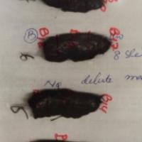
巨检:灰褐色组织9.5x4x2.5cm,切面囊实性,实性区灰白灰褐质中,
囊性区见清澄色液体。
镜检:切片见成熟的复磷上皮、假复层纤毛柱状上皮、纤维及血管组织,局灶区见神经组织。
IHC15-306
CK(+)EMA(+)Cam5.2(+)vim(+)CD34(+)
CD45(+)SMA(+)MSA(+)Syn(+)s-100(+)
Des(—)Ki67(+)(5%)
病理诊断:纵隔成熟畸胎瘤,局灶区见神经组织,请临床注意随访。
-
蝴蝶兰dali2003 离线
- 帖子:11456
- 粉蓝豆:2441
- 经验:12021
- 注册时间:2009-09-06
- 加关注 | 发消息


