共2页/36条首页上一页12下一页尾页
回复:39 阅读:5956
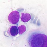
解释一下:“国外病例,稍后公布答案:Ascites cytology, 34-year old female,positive for adenocarcinoma

junzi003981 离线

帖子:1469
粉蓝豆:234
经验:3249
注册时间:2008-09-07
加关注  |  发消息
楼主 发表于 2013-05-05 17:33|举报|关注(3)
浏览排序[ 顺序 逆序 楼主 支持 精彩 ]  快捷回复
性别年龄34
一般病史none
标本类型Ascites fluid
制片方法none
染色方法Romanovsky stains

  • 解释一下:“国外病例,稍后公布答案:Ascites cytology, 34-year old female,positive for adenocarcinoma图1
    图1
  • 解释一下:“国外病例,稍后公布答案:Ascites cytology, 34-year old female,positive for adenocarcinoma图2
    图2
  • 解释一下:“国外病例,稍后公布答案:Ascites cytology, 34-year old female,positive for adenocarcinoma图3
    图3
  • 解释一下:“国外病例,稍后公布答案:Ascites cytology, 34-year old female,positive for adenocarcinoma图4
    图4
  • 解释一下:“国外病例,稍后公布答案:Ascites cytology, 34-year old female,positive for adenocarcinoma图5
    图5


标签:病例
本帖最后由 junzi003981 于 2013-05-12 21:45:34 编辑
0
signature
为人类医疗事业贡献一生
添加参考诊断
×参考诊断
  

旅顺病理 离线

帖子:4084
粉蓝豆:144
经验:5113
注册时间:2011-06-04
加关注  |  发消息
2 楼    发表于2013-05-06 11:18:41举报|引用
返回顶部 | 快捷回复

“毛”是哪来的

细胞也有问题

2

竹林心语

ma830120
收起回复

et666 离线

帖子:386
粉蓝豆:18
经验:658
注册时间:2008-03-11
加关注  |  发消息
20 楼    发表于2013-11-23 20:16:30举报|引用
返回顶部 | 快捷回复

 腹水中细胞团有立体感、细胞异型性明显,有清晰的核仁,有分泌空泡 ,符合腺癌特征。    

 谢谢楼主为大家分享了这么好的病例!

1

文长江
回复
signature
王秀娥

gwls0008 离线

帖子:1365
粉蓝豆:5
经验:1465
注册时间:2008-08-24
加关注  |  发消息
1 楼    发表于2013-05-05 18:13:17举报|引用
返回顶部 | 快捷回复

反应性间皮。

1

islguo
回复

junzi003981 离线

帖子:1469
粉蓝豆:234
经验:3249
注册时间:2008-09-07
加关注  |  发消息
11 楼    发表于2013-05-12 00:04:17举报|引用
返回顶部 | 快捷回复
本帖最后由 junzi003981 于 2013-05-12 00:10:50 编辑

  我看了大量国外和国内资料,给大家总结一下:

微小绒毛可见良性间皮细胞、间皮瘤、卵巢癌、乳腺癌和胃癌细胞。但是,卵巢癌肿瘤细胞微绒毛长度一般分布长短不一,比良性间皮细胞微绒毛更长,通常出现在细胞一极,也叫假绒毛,但是也可能整个细胞覆着微绒毛,微绒毛也可以出现在乳腺癌、间皮瘤和胃癌细胞,但是腹水中的卵巢癌细胞常见。巴氏染色很难看到微绒毛,巴格氏染色(Romanowsky Stained )和瑞氏吉姆萨染色更能清楚可见了,近来有报道糖原染色也可见微绒毛。但是需要提醒大家,是恶性还是良性,需要结合细胞核和胞浆特点来判断。

  • 图1
  • 图2
  • 图3
  • 图4
  • 图5
  • 图6
  • 图7
  • 图8
  • 图9
  • 图10

1

ruibo
回复
signature
为人类医疗事业贡献一生

gwls0008 离线

帖子:1365
粉蓝豆:5
经验:1465
注册时间:2008-08-24
加关注  |  发消息
10 楼    发表于2013-05-11 18:53:08举报|引用
返回顶部 | 快捷回复

为什么就不是反应性间皮呢?而一定是肿瘤?????老外只是说不是恶性间皮瘤。而在卵巢有“浆液性乳头状囊腺癌”。说腹水中出现的细胞就是癌细胞,无形态学依据,无免疫组化依据。it's pointless and baseless.

1

月落123
回复

gwls0008 离线

帖子:1365
粉蓝豆:5
经验:1465
注册时间:2008-08-24
加关注  |  发消息
16 楼    发表于2013-05-17 18:47:48举报|引用
返回顶部 | 快捷回复

认真复习过后,仍感觉细胞形态有重叠,不能根据一个诊断标准进行诊断。

0
回复

fuyong 离线

帖子:6879
粉蓝豆:1938
经验:12692
注册时间:2008-03-10
加关注  |  发消息
5 楼    发表于2013-05-06 12:09:31举报|引用
返回顶部 | 快捷回复

增生间皮或间皮瘤

0
回复
signature
多读书,读好书

xg120 离线

帖子:12586
粉蓝豆:1205
经验:15200
注册时间:2011-10-14
加关注  |  发消息
6 楼    发表于2013-05-06 14:22:59举报|引用
返回顶部 | 快捷回复

增生间皮细胞?或间皮瘤

0
回复
signature
尚泽忠

ahdj 离线

帖子:17
粉蓝豆:11
经验:69
注册时间:2012-07-04
加关注  |  发消息
7 楼    发表于2013-05-06 15:06:48举报|引用
返回顶部 | 快捷回复

间皮来源肿瘤?

0
回复

路过蜻蜓 离线

帖子:756
粉蓝豆:748
经验:1057
注册时间:2010-09-03
加关注  |  发消息
15 楼    发表于2013-05-13 23:34:22举报|引用
返回顶部 | 快捷回复

 腹水中见细胞异型性明显,有清晰的核仁,有分泌空泡+微绒毛(比良性间皮细胞微绒毛更长),考虑卵巢癌,是这个意思吧?谢谢楼主为大家分享了这么好的病例!

0
回复
signature
一天一点小进步!

yy2010 离线

帖子:123
粉蓝豆:27
经验:169
注册时间:2010-11-04
加关注  |  发消息
14 楼    发表于2013-05-13 10:47:31举报|引用
返回顶部 | 快捷回复


0
回复
signature
在学习中成长。

月落123 离线

帖子:132
粉蓝豆:6
经验:220
注册时间:2012-05-31
加关注  |  发消息
18 楼    发表于2013-10-30 10:08:47举报|引用
返回顶部 | 快捷回复

这个腹水的细胞,看上去是间皮细胞,并不是说是卵巢癌,腹水就一定有癌细胞

0
回复

ahdj 离线

帖子:17
粉蓝豆:11
经验:69
注册时间:2012-07-04
加关注  |  发消息
12 楼    发表于2013-05-12 23:25:05举报|引用
返回顶部 | 快捷回复

 学习了

0
回复

zwx 离线

帖子:3
粉蓝豆:1
经验:14
注册时间:2011-08-14
加关注  |  发消息
16 楼    发表于2013-09-03 21:46:22举报|引用
返回顶部 | 快捷回复

学习!

0
回复

zangwuying 离线

帖子:280
粉蓝豆:73
经验:685
注册时间:2012-07-28
加关注  |  发消息
17 楼    发表于2013-09-04 13:06:28举报|引用
返回顶部 | 快捷回复

细胞病理在不断总结中前进,谢谢楼主总结的这一条经验

0
回复
signature
求真求实

godwind 离线

帖子:129
粉蓝豆:206
经验:980
注册时间:2015-12-23
加关注  |  发消息
36 楼    发表于2017-03-13 13:26:10举报|引用
来自iPhone 返回顶部 | 快捷回复
学习了
0
回复

旅顺病理 离线

帖子:4084
粉蓝豆:144
经验:5113
注册时间:2011-06-04
加关注  |  发消息
9 楼    发表于2013-05-09 11:14:18举报|引用
返回顶部 | 快捷回复

0
回复

蓝宝石6628 离线

帖子:16787
粉蓝豆:321
经验:17245
注册时间:2012-03-30
加关注  |  发消息
13 楼    发表于2013-05-13 06:01:14举报|引用
返回顶部 | 快捷回复

0
回复
signature
乐观向上,不断进取!

junzi003981 离线

帖子:1469
粉蓝豆:234
经验:3249
注册时间:2008-09-07
加关注  |  发消息
8 楼    发表于2013-05-08 21:39:29举报|引用
返回顶部 | 快捷回复
本帖最后由 junzi003981 于 2013-05-08 21:43:54 编辑
    • 图1
    • 图2
    • 图3
    • 图4

    卵巢乳头状癌 Papillary carcinoma of ovary


    Anemone cells: the presence of many microvilli in malignant cells is frequently documented by electron microscopy (EM), but their presence can be seen in some cases by optical microscopy as well. The use of Romanovsky stains can be very useful in this context ( I use to say they are the EM of the poor...). Spriggs and Boddington, in their classic book Cytology of effusions, Grune & Stratton, 1968, 2nd edition, were the first to call attention to this finding in papillary carcinomas of the ovary, with pictures remarkably similar to the ones in our cases. More recently, this finding has been called "anemone cells" and "anemone tumors" in relation to the cells or the tumors bearing them, respectively. 
    This case illustrates the difficult differential diagnosis between this type of tumor and mesothelioma, in special of the surface of the ovary and the pelvic peritoneum. Both tumors may exhibit windows between the cells, and microvilli in their surface. There is no immunochemistry test of real value, and the only certainty is done by the surgical exploration, as done in this case, where the pelvic peritoneum was free of disease, and the ovary had a typical serous cystadenocarcinoma papillary tumor. Their coelomic epithelial common origin explains their extreme similarities. Sometimes they both have ciliated cells besides microvilli in their surface.

0
回复
signature
为人类医疗事业贡献一生

scj1233 离线

帖子:383
粉蓝豆:13
经验:405
注册时间:2010-11-06
加关注  |  发消息
19 楼    发表于2013-11-09 20:05:39举报|引用
返回顶部 | 快捷回复

 只看图片会考虑恶性间皮瘤。

0
回复
回复:39 阅读:5956
共2页/36条首页上一页12下一页尾页
【免责声明】讨论内容仅作学术交流之用,不作为诊疗依据,由此而引起的法律问题作者及本站不承担任何责任。
快速回复
进入高级回复
您最多可输入10000个汉字,按 "Ctrl" + "Enter" 直接发送
搜索回复/乘电梯 ×
按内容
按会员
乘电梯
合作伙伴
友情链接