


| 图片: | |
|---|---|
| 名称: | |
| 描述: | |
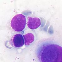
- 解释一下:“国外病例,稍后公布答案:Ascites cytology, 34-year old female,positive for adenocarcinoma
-
junzi003981 离线
- 帖子:1469
- 粉蓝豆:234
- 经验:3249
- 注册时间:2008-09-07
- 加关注 | 发消息
-
junzi003981 离线
- 帖子:1469
- 粉蓝豆:234
- 经验:3249
- 注册时间:2008-09-07
- 加关注 | 发消息
-
本帖最后由 junzi003981 于 2013-05-08 21:43:54 编辑
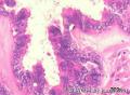 图1
图1 图2
图2 图3
图3 图4
图4
卵巢乳头状癌 Papillary carcinoma of ovary
Anemone cells: the presence of many microvilli in malignant cells is frequently documented by electron microscopy (EM), but their presence can be seen in some cases by optical microscopy as well. The use of Romanovsky stains can be very useful in this context ( I use to say they are the EM of the poor...). Spriggs and Boddington, in their classic book Cytology of effusions, Grune & Stratton, 1968, 2nd edition, were the first to call attention to this finding in papillary carcinomas of the ovary, with pictures remarkably similar to the ones in our cases. More recently, this finding has been called "anemone cells" and "anemone tumors" in relation to the cells or the tumors bearing them, respectively.
This case illustrates the difficult differential diagnosis between this type of tumor and mesothelioma, in special of the surface of the ovary and the pelvic peritoneum. Both tumors may exhibit windows between the cells, and microvilli in their surface. There is no immunochemistry test of real value, and the only certainty is done by the surgical exploration, as done in this case, where the pelvic peritoneum was free of disease, and the ovary had a typical serous cystadenocarcinoma papillary tumor. Their coelomic epithelial common origin explains their extreme similarities. Sometimes they both have ciliated cells besides microvilli in their surface.

- 为人类医疗事业贡献一生
-
junzi003981 离线
- 帖子:1469
- 粉蓝豆:234
- 经验:3249
- 注册时间:2008-09-07
- 加关注 | 发消息
-
本帖最后由 junzi003981 于 2013-05-12 00:10:50 编辑
 图1
图1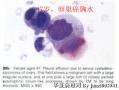 图2
图2 图3
图3 图4
图4 图5
图5 图6
图6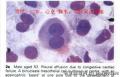 图7
图7 图8
图8 图9
图9 图10
图10
我看了大量国外和国内资料,给大家总结一下:
微小绒毛可见良性间皮细胞、间皮瘤、卵巢癌、乳腺癌和胃癌细胞。但是,卵巢癌肿瘤细胞微绒毛长度一般分布长短不一,比良性间皮细胞微绒毛更长,通常出现在细胞一极,也叫假绒毛,但是也可能整个细胞覆着微绒毛,微绒毛也可以出现在乳腺癌、间皮瘤和胃癌细胞,但是腹水中的卵巢癌细胞常见。巴氏染色很难看到微绒毛,巴格氏染色(Romanowsky Stained )和瑞氏吉姆萨染色更能清楚可见了,近来有报道糖原染色也可见微绒毛。但是需要提醒大家,是恶性还是良性,需要结合细胞核和胞浆特点来判断。
- 为人类医疗事业贡献一生
-
zangwuying 离线

- 帖子:280
- 粉蓝豆:73
- 经验:685
- 注册时间:2012-07-28
- 加关注 | 发消息


