共2页/22条首页上一页12下一页尾页
回复:22 阅读:8206
左上腹肿块

zhanglei 离线

帖子:3751
粉蓝豆:2078
经验:3901
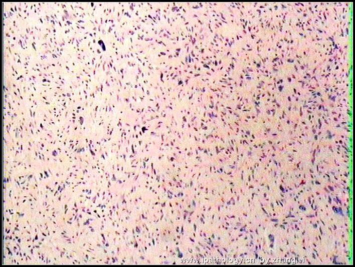
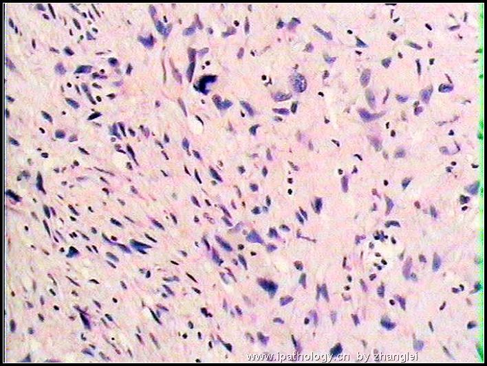
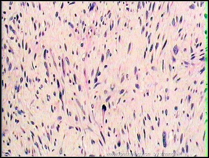
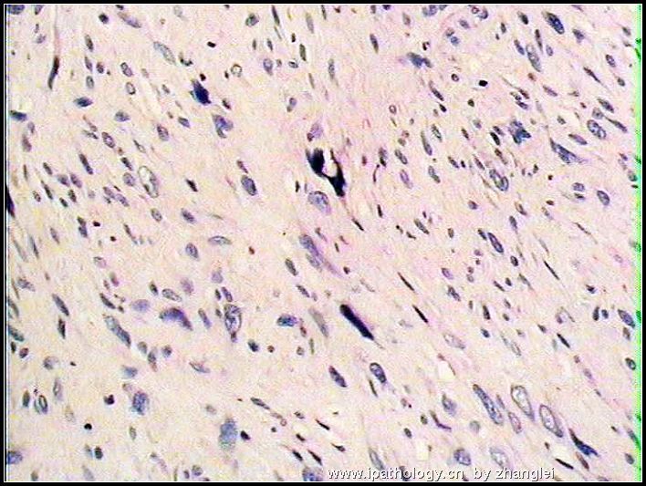
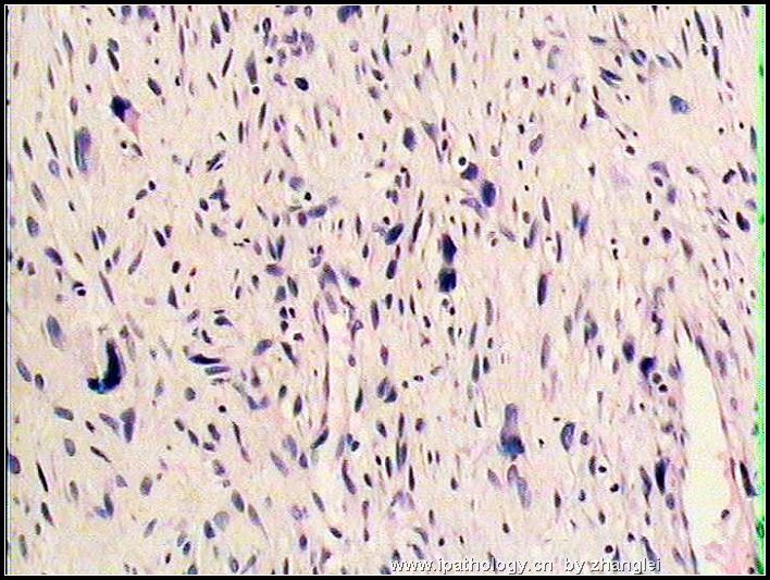
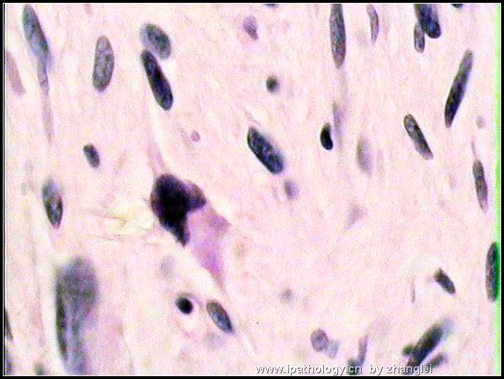
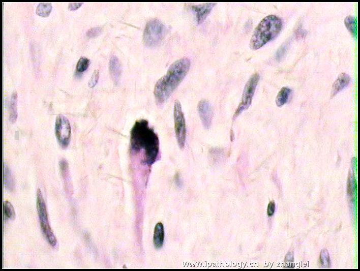
注册时间:2006-09-26
加关注  |  发消息
楼主 发表于 2006-11-12 21:28|举报|关注(1)
浏览排序[ 顺序 逆序 楼主 支持 精彩 ]  快捷回复
姓    名: ××× 性别:   年龄:  44
标本名称:  左上腹肿块
简要病史:  发现左上腹肿块一周。术中见左肾下极巨大肿块25*20*10cm
肉眼检查: 不整形组织一块18*14*9cm,切面灰白色,质韧,肿块与肾下极粘连并压迫肾脏。
左上腹肿块图1
名称:图1
描述:图1
左上腹肿块图2
名称:图2
描述:图2
左上腹肿块图3
名称:图3
描述:图3
左上腹肿块图4
名称:图4
描述:图4
左上腹肿块图5
名称:图5
描述:图5
左上腹肿块图6
名称:图6
描述:图6
左上腹肿块图7
名称:图7
描述:图7
标签:腹膜后 低度恶性神经鞘膜瘤 脂肪肉瘤
本帖最后由 于 2006-11-12 23:17:00 编辑
0
×参考诊断
上海肿瘤医院会诊:低度恶性神经鞘膜瘤

安迪 离线

帖子:598
粉蓝豆:3525
经验:849
注册时间:2011-03-04
加关注  |  发消息
1 楼    发表于2011-06-09 19:18:00举报|引用
返回顶部 | 快捷回复
 学习
0
回复

197 离线

帖子:4781
粉蓝豆:803
经验:5032
注册时间:2006-09-12
加关注  |  发消息
2 楼    发表于2006-11-12 22:34:00举报|引用
返回顶部 | 快捷回复
本帖最后由 于 2006-11-12 22:34:00 编辑 看起来似有病理性分裂相。
和子宫的关系如何?子宫的情况如何?
多取块,看看有没有脂肪的成份,以便鉴别血管平滑肌脂肪瘤。
看这几张图,首先想到三个病:平滑肌肉瘤,间质瘤,恶性纤维组织细胞瘤。
数数分裂相,仔细逐条核对平滑肌肉瘤的诊断标准。
做几项免疫组化,帮助诊断和鉴别:Vim, Des,MSA,CD117, CD34,S-100, NSE.CD68, 溶菌酶
0
回复
signature
“人生没有彩排,每一天都是现场直播”

shihuaiy 离线

帖子:707
粉蓝豆:35
经验:707
注册时间:2006-09-27
加关注  |  发消息
3 楼    发表于2006-11-12 23:05:00举报|引用
返回顶部 | 快捷回复
此例很有意思,欢迎大家继续讨论。
0
回复
signature
the more we discuss, the more we learn from each other !!

mjma 离线

帖子:703
粉蓝豆:24
经验:789
注册时间:2006-09-28
加关注  |  发消息
4 楼    发表于2006-11-13 01:56:00举报|引用
返回顶部 | 快捷回复
A retroperitoneal tumor of such large size and histology (spindled cells) has to raise the possibility of dedifferentiated liposarcoma. I urge that any "fat" attached to the tumor be submitted for microscopic examination to detect any feature for a pre-existing and residual low grade liposarcoma. Retroperitoneal liposarcomas are not rare, and are the most common large retroperitoneal tumors in adults. Most of them are low grade or well differentiated liposarcoma (rather than the myxoid or pleomorphic variants of liposarcomas). Microscopically, retroperitoneal well differentiated liposarcomas appear identical to the well circumscribed atypical lipomatous tumors (atypical lipomas) of neck, shoulder, upper back and other regions in extremities. However, retroperitoneal well-differentiated liposarcomas are slow-growing, very large, not encapsulated and very difficult to resect completely due to infiltrative borders.

A minority of retroperitoneal liposarcomas may, either at the time of initial diagnosis or at the time of local recurrence, contain nodule(s) of non-lipogenic sarcomatous elements. Most of these non-lipogenic elements appear like fibrosarcoma or MFH, and often display features of high grade sarcoma. Sometimes, the sarcomatous elements are that of a low grade fibrosarcoma (consisting exclusively of spindled cells). Very rarely, features of high grade osteosarcoma, leiomyosarcoma, chondrosarcoma, rhabdomyosarcoma and malignant peripheral nerve sheath tumor are found. These tumors are known as dedifferentiated liposarcomas. They are presumed to evolve from the pre-existing well differentiated liposarcomas. Unlike well differentiated liposarcomas, dedifferentiated liposarcomas of the retroperitoneum have definite metastatic potential (in 1~18% of cases). No matter what kinds of sarcomatous elements are found in a retroperitoneal dedifferentiated liposarcoma, its clinical behavior is not the same as high grade fibrosarcomas, leiomyosarcomas, osteosarcomas, chondrosarcomas, rhabdomyosarcomas, or MPNST. Dedifferentiated liposarcomas are not unique to the retroperitoneum. Another common location for it is the inguinal area and spermatic cord.
0
回复
signature

聞道有先後,術業有專攻

zhanglei 离线

帖子:3751
粉蓝豆:2078
经验:3901
注册时间:2006-09-26
加关注  |  发消息
5 楼    发表于2007-02-04 13:15:00举报|引用
返回顶部 | 快捷回复
以下是引用怀江 在2006-12-13 19:26:00的发言:

以下是引用zhanglei 在2006-12-2 0:46:00的发言:

CD34++  八因子+  CEA  HMB45  S-100+      上海肿瘤医院会诊:低度恶性神经鞘膜瘤。

就这些免疫组画排除不了去分化脂肪肉瘤.不如做个脂肪染色看看

读片会的资料,此例讨论至此,我想明确最后诊断结果已经不是很重要了,因为从各位老师的讨论中,我已经学到了很多。。。。
0
回复

tianxin 离线

帖子:31
粉蓝豆:1
经验:31
注册时间:2006-09-25
加关注  |  发消息
6 楼    发表于2006-11-14 23:22:00举报|引用
返回顶部 | 快捷回复
以下是引用mjma 在2006-11-13 1:56:00的发言:

A retroperitoneal tumor of such large size and histology (spindled cells) has to raise the possibility of dedifferentiated liposarcoma. I urge that any "fat" attached to the tumor be submitted for microscopic examination to detect any feature for a pre-existing and residual low grade liposarcoma. Retroperitoneal liposarcomas are not rare, and are the most common large retroperitoneal tumors in adults. Most of them are low grade or well differentiated liposarcoma (rather than the myxoid or pleomorphic variants of liposarcomas). Microscopically, retroperitoneal well differentiated liposarcomas appear identical to the well circumscribed atypical lipomatous tumors (atypical lipomas) of neck, shoulder, upper back and other regions in extremities. However, retroperitoneal well-differentiated liposarcomas are slow-growing, very large, not encapsulated and very difficult to resect completely due to infiltrative borders.

A minority of retroperitoneal liposarcomas may, either at the time of initial diagnosis or at the time of local recurrence, contain nodule(s) of non-lipogenic sarcomatous elements. Most of these non-lipogenic elements appear like fibrosarcoma or MFH, and often display features of high grade sarcoma. Sometimes, the sarcomatous elements are that of a low grade fibrosarcoma (consisting exclusively of spindled cells). Very rarely, features of high grade osteosarcoma, leiomyosarcoma, chondrosarcoma, rhabdomyosarcoma and malignant peripheral nerve sheath tumor are found. These tumors are known as dedifferentiated liposarcomas. They are presumed to evolve from the pre-existing well differentiated liposarcomas. Unlike well differentiated liposarcomas, dedifferentiated liposarcomas of the retroperitoneum have definite metastatic potential (in 1~18% of cases). No matter what kinds of sarcomatous elements are found in a retroperitoneal dedifferentiated liposarcoma, its clinical behavior is not the same as high grade fibrosarcomas, leiomyosarcomas, osteosarcomas, chondrosarcomas, rhabdomyosarcomas, or MPNST. Dedifferentiated liposarcomas are not unique to the retroperitoneum. Another common location for it is the inguinal area and spermatic cord.

      腹膜后肿瘤,具有如此体积和组织学形态(梭形细胞)让我们不得不考虑去分化脂肪肉瘤的可能性。我强烈建议将任何与该肿瘤相连的脂肪组织送做镜检以发现已有和残留的低级别脂肪肉瘤的特征。腹膜后脂肪肉瘤并不罕见,是成人腹膜后肿瘤中的一个最常见类型。大部分都属于低级别或分化良好型脂肪肉瘤(而非粘液型或多形性脂肪肉瘤)。显微镜下,腹膜后分化良好型脂肪肉瘤在形态上与发生在颈、肩、上背和四肢其他部位的境界清楚的非典型脂肪瘤相似。但是腹膜后分化良好型脂肪肉瘤生长缓慢,体积可以很大,无包膜,由于边界不清很难切除干净。
      少数腹膜后脂肪肉瘤可以在初次诊断或局部复发时发现局灶区非脂肪肉瘤的肉瘤成分。大部分非脂肪肉瘤成分镜下很像纤维肉瘤或恶性纤维组织细胞瘤,并且通常显示的是高级别肉瘤的特征。罕见的情况下可以找见高级别骨肉瘤、平滑肌肉瘤、软骨肉瘤、横纹肌肉瘤和恶性外周神经鞘膜瘤的特征。这样的肿瘤被称为去分化的脂肪肉瘤。它们被认为是在已有的分化良好型脂肪肉瘤的基础上发展而来的。与分化良好型脂肪肉瘤不同,腹膜后的去分化脂肪肉瘤具有一定的转移倾向(占病例数的1-18%)。无论在腹膜后去分化型脂肪肉瘤中发现何种类型的其他肉瘤成分,它的临床行为都与高级别纤维肉瘤、平滑肌肉瘤、骨肉瘤、软骨肉瘤、或恶性外周神经鞘膜瘤不同。腹膜后并不是去分化脂肪肉瘤发生的唯一部位。其他常见部位包括腹股沟区和精索。
0
回复

shihuaiy 离线

帖子:707
粉蓝豆:35
经验:707
注册时间:2006-09-27
加关注  |  发消息
7 楼    发表于2006-11-15 23:31:00举报|引用
返回顶部 | 快捷回复

感觉此例有四个主要形态特点有助于诊断:1 第一图的storiform排列;2 背景稀疏,有些粘液样改变;3 部分细胞核染色质非常深染如墨水点样;4 个别细胞胞浆丰富,核偏位,似脂母细胞(最后一张图所示)。

0
回复
signature
the more we discuss, the more we learn from each other !!

心雨 离线

帖子:419
粉蓝豆:1
经验:687
注册时间:2011-04-17
加关注  |  发消息
8 楼    发表于2011-04-23 12:12:00举报|引用
返回顶部 | 快捷回复
以下是引用zhanglei 在2006-12-2 0:46:00的发言:


CD34++  八因子+  CEA  HMB45  S-100+      上海肿瘤医院会诊:低度恶性神经鞘膜瘤。

学习
0
回复

llb-1 离线

帖子:76
粉蓝豆:1
经验:120
注册时间:2006-10-07
加关注  |  发消息
9 楼    发表于2007-02-05 23:07:00举报|引用
返回顶部 | 快捷回复
 我想明确最后诊断结果已经不是很重要了,因为从各位老师的讨论中,我已经学到了很多。。。。
0
回复

慧眼 离线

帖子:98
粉蓝豆:2
经验:98
注册时间:2006-11-17
加关注  |  发消息
10 楼    发表于2006-11-18 22:51:00举报|引用
返回顶部 | 快捷回复
恶性外周神经鞘膜瘤。
0
回复

qwe628400 离线

帖子:459
粉蓝豆:57
经验:516
注册时间:2009-11-17
加关注  |  发消息
11 楼    发表于2011-06-15 20:47:00举报|引用
返回顶部 | 快捷回复
 dedifferentiated liposarcoma
0
回复

zhanglei 离线

帖子:3751
粉蓝豆:2078
经验:3901
注册时间:2006-09-26
加关注  |  发消息
12 楼    发表于2006-12-02 12:46:00举报|引用
返回顶部 | 快捷回复
 

CD34++  八因子+  CEA  HMB45  S-100+      上海肿瘤医院会诊:低度恶性神经鞘膜瘤。

0
回复

爱兰 离线

帖子:380
粉蓝豆:4
经验:388
注册时间:2006-10-02
加关注  |  发消息
13 楼    发表于2006-12-02 11:30:00举报|引用
返回顶部 | 快捷回复

 从你发的图象看有栅栏样排列.,细胞呈梭形大小不一.,大部分细胞比较温和,核分裂像不多见.有奇异核.

腹膜后肿瘤的特点发现比较晚,由于空间大肿瘤一般都很大.腹膜后易发生神经源性肿瘤要与去分化脂肪肉瘤鉴别..

分化脂肪肉瘤梭形成分比这个要恶像纤维肉瘤样改变.

0
回复
signature
金杯银杯不如人们的口杯

shihuaiy 离线

帖子:707
粉蓝豆:35
经验:707
注册时间:2006-09-27
加关注  |  发消息
14 楼    发表于2006-12-02 16:37:00举报|引用
返回顶部 | 快捷回复

非常同意 马教授的观点,此例从细胞排列、形态以及间质特点来看,都需要首先考虑去分化脂肪肉瘤的可能。免疫组化在区分去分化脂肪肉瘤与外周神经肿瘤方面好像并没有太大帮助,多取材找相对典型区域可能帮助更大。最近有文献报道MDM-2以及CDK-4在去分化脂肪肉瘤阳性率较高,我们用的结果也还可以。就此例形态来讲主要是去分化脂肪肉瘤、MFH、MPNST之间的鉴别,但有文献报道腹膜后MFH多数可能都是去分化脂肪肉瘤,如果此结论可信的话,这例主要是去分化脂肪肉瘤和MPNST之间的鉴别了。

建议再多取材找找有无相对典型一些的区域。再追问一下以往有无手术史,如果有的话可以复查一下原切片。

0
回复
signature
the more we discuss, the more we learn from each other !!

spellq 离线

帖子:134
粉蓝豆:23
经验:150
注册时间:2006-09-27
加关注  |  发消息
15 楼    发表于2006-12-05 19:40:00举报|引用
返回顶部 | 快捷回复
 上次有个病例,女性,52岁,发生在肾脏的一个肿块。大体:肿块充满整个肾脏,直径16cm左右,切面灰白色,结节状。镜下:见大片状坏死和粘液,粘液中漂浮着梭形的细胞,还有的区域甚至出现storiform结构,核分裂多见。后来原单位发了:粘液型脂肪肉瘤
0
回复
signature
三人行,必有我师焉!

yifan 离线

帖子:100
粉蓝豆:1
经验:100
注册时间:2006-11-18
加关注  |  发消息
16 楼    发表于2006-12-11 21:14:00举报|引用
返回顶部 | 快捷回复
 非常喜欢zhanglei这样的发图方式,看起来方便
0
回复

197 离线

帖子:4781
粉蓝豆:803
经验:5032
注册时间:2006-09-12
加关注  |  发消息
17 楼    发表于2006-12-11 23:09:00举报|引用
返回顶部 | 快捷回复
 
0
回复
signature
“人生没有彩排,每一天都是现场直播”

Chiang 离线

帖子:2443
粉蓝豆:1085
经验:3336
注册时间:2006-10-03
加关注  |  发消息
18 楼    发表于2006-12-12 08:50:00举报|引用
返回顶部 | 快捷回复
 发生于腹膜后类似结构的肿瘤多半是去分化脂肪肉瘤,有时候脂肪成分可能很少,甚至见不到脂肪成分,尤其是出现所谓的恶性纤维组织细胞瘤结构时,恶纤组的诊断要慎重,或尽量少诊断。
0
回复

JX16 离线

帖子:1079
粉蓝豆:5
经验:3225
注册时间:2008-10-27
加关注  |  发消息
19 楼    发表于2011-06-01 20:39:00举报|引用
返回顶部 | 快捷回复
 学习
0
回复
signature
jx16

怀江 离线

帖子:20
粉蓝豆:2
经验:34
注册时间:2006-11-01
加关注  |  发消息
20 楼    发表于2006-12-13 19:26:00举报|引用
返回顶部 | 快捷回复
以下是引用zhanglei 在2006-12-2 0:46:00的发言:

CD34++  八因子+  CEA  HMB45  S-100+      上海肿瘤医院会诊:低度恶性神经鞘膜瘤。

就这些免疫组画排除不了去分化脂肪肉瘤.不如做个脂肪染色看看
0
回复
回复:22 阅读:8206
共2页/22条首页上一页12下一页尾页
【免责声明】讨论内容仅作学术交流之用,不作为诊疗依据,由此而引起的法律问题作者及本站不承担任何责任。
快速回复
进入高级回复
您最多可输入10000个汉字,按 "Ctrl" + "Enter" 直接发送
搜索回复/乘电梯 ×
按内容
按会员
乘电梯
合作伙伴
友情链接