| 图片: | |
|---|---|
| 名称: | |
| 描述: | |
- 乳腺肿块
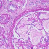
Generally 间质内的腺体没有显示肌上皮 suggest invasive ca. However, you must make sure your stains were good. If this case is a papillary lesion and these glands are within the sclerosing stroma within the papillary lesion, the chance of invasion is low. Anyway I am confused for this case and cannot make the decision based on the photos
谢谢赵老师,根据以往经验和此次内对照显示标记应该是可靠的,因此这个病例我们也非常困惑,因为患者才31岁,而且没有生育,鉴于此,我们建议临床医生扩大切除即可,临床医师反馈肿块位于乳晕下方,无法扩大切除,所以只能建议会诊,上级医院会诊考虑旺炽性导管内乳头状瘤,建议密切随访!
再次感谢赵老师不厌其烦的指导!
Generally 间质内的腺体没有显示肌上皮 suggest invasive ca. However, you must make sure your stains were good. If this case is a papillary lesion and these glands are within the sclerosing stroma within the papillary lesion, the chance of invasion is low. Anyway I am confused for this case and cannot make the decision based on the photos
-
shihong4699 离线
- 帖子:1024
- 粉蓝豆:43
- 经验:2917
- 注册时间:2009-01-20
- 加关注 | 发消息
Papillary lesions show areas without myoepithelial cells and the areas with myoepithelial cells.
Papillary DCIS or atypical papilloma.?
It should be easy for this case if you read true slides of H&E, and IHC.
Cytologic features are not bad or not like cancer.
Maybe atypical papilloma.
?????
非常感谢!
Papillary lesions show areas without myoepithelial cells and the areas with myoepithelial cells.
Papillary DCIS or atypical papilloma.?
It should be easy for this case if you read true slides of H&E, and IHC.
Cytologic features are not bad or not like cancer.
Maybe atypical papilloma.
?????


