


 哈哈! 明白了.


| 图片: | |
|---|---|
| 名称: | |
| 描述: | |
- (推荐)Askin 瘤的起源及本质
Askin 瘤的起源及本质
范钦和 南京医科大学一附院 210029
摘要 目的:探讨Askin 瘤的本质,起源。方法:以临床—病理观察,免疫组织化学,电子显微镜技术和细胞遗传技术对22例Askin瘤进行研究。结果:显示瘤细胞免疫组化为MIC2(CD99),NSE、cgA 阳性;电镜见神经分泌颗粒;细胞遗传分析示特异性基因遗传异位,即t(11;22)(q24;q12)。这些特点与恶性原始神经外胚叶肿瘤(PNET)相同。结论:我们认为Askin瘤实际上是PNET/尤文氏肉瘤家族的一员,只是发生部位较特殊,预后不良。应与其它小圆细胞肉瘤如淋巴瘤,胚胎型横纹肌肉瘤,横纹肌样瘤,圆细胞脂肪肉瘤,结缔组织生成性小圆细胞肿瘤相鉴别。
关键词 Askin瘤;PNET/尤文氏肉瘤;免疫组化;电子显微镜;
细胞遗传分型
中国图书分类号 R:738.6
作者单位:南京医科大学第一附属医院理科210029
作者简介:范钦和,男,48岁,硕士研究生毕业,主任医师,教授,病理科主任。国际外科病理学杂志编委,澳太地区软组织肿瘤会诊中心委员,江苏省病理学会和肿瘤学会主任委员。研究方向:软组织肿瘤
The origination and reality of Askin tumor
1Fan Qinhe, 2Philip W. Allen, 1Xu Tianrong et al
(1Department of Pathology, The First Affiliated Hospital of Nanjing Medical University, Nanjing 210029)
ABSTRACT Purpose To study the origination and reality of Askin tumor. Method Twenty-two cases of these lesions have been studied by clinical-pathologic observation, immunohistochemistry, electron-microscopy, and cytogenics in this paper. Results The results revealed the positiveness of MIC2(CD99), NSE, Chromogranin A(CgA) by immunohistochemistry, neurosecretory granules by electron-microscopy, and the specific genotypic translocation of t(11:22)(q24:q12) by cytogenetic analysis, which were identical to the malignant primitive neuroectodermal tumor (PNET). Conclusion We conclude that this lesion is actually the one member of the PNET/Ewing’s sarcoma family, except the special location. Differential diagnoses between this tumor and other small round cell sarcoma such as lymphoma, embryonal rhabdomyosarcoma, rhabdoid tumor, round cell liposarcoma and desmoplastic small round cell tumor were also discussed in this article.
KEY WORDS Askin tumor; PNET/Ewing’s sarcoma; immunohistochemistry; electron-microscopy; Cytogenetic analysis
1979年,Askin等人首先描述了20例“儿童和青少年胸肺部恶性小细胞肿瘤”,认为其有别於尤文氏肉瘤,神经母细胞瘤,神经上皮瘤,胚胎型横纹肌肉瘤,淋巴瘤等小细胞性肉瘤,是一种起源末定的新的独立类型(1),其后,不少作者也报告了各自的类似发现,并称之为Askin瘤(2、3)。
Askin瘤可见於胸壁软组织,肋骨骨膜、肺。瘤组织主要由大小较一致的小圆细胞所组成,排列成片块状或形成假小叶。肿瘤易复发和转移,为高度恶性、临床预后差。
随着免疫组化、电子显微镜及分子生物学技术的发展,人们对Askin 瘤有了深入的认识。本文作者就自己所收集的确22例Askin 瘤进行临床--病理、免疫组化、电镜状和细胞遗传学研究,以进一步探讨其起源和本质。
1 材料及方法
22例Askin瘤来自江苏省人民医院(南京医科大学一附院)病理科和澳太地区软组织肿瘤会诊中心1985-1998共14年间的常规活检病例和会诊病例。原诊时名称为Askin 瘤和胸(肺)部恶性小园细胞肿瘤。
所有病例均以福尔马林固定,常规处理。复习了所有H.E切片,初步确认诊断。其中18例做了免疫组织化学,第一抗体有MIC2(CD99),神经元特异性烯醇化酶(NSE),突触素(Synaptophysin),嗜铬蛋白A(Chromogranin A), S-100蛋白,胶质原纤维酸性蛋白(GFAP),波形纤维蛋白(Vimentin),角蛋白(Keratin),白细胞共同抗原(LCA)。采取ABC、PAP法。试剂分别来自DAKO公司,Vector公司和Sigma公司。
5例做了电子显微镜观察,取新鲜肿瘤组织标本固定於戌二醛,按电镜标本制作程序制作成铜网后上透射电子显微镜观察。
4例作了细胞遗传学检查,均采取新鲜肿瘤组织标本由细胞遗传学实验室作染色体核型分析。
2 临床资料
患者以儿童和青少年为主,年龄范围1-23岁,平均年龄13岁。男:女为12:10,其中白人10例,亚洲人12例。病人主要症状和体征依次为胸壁肿块,胸痛,呼吸困难,发热和胸水。肿瘤发生部位依次为胸壁软组织(常累及肋骨)18例;壁层和脏层胸膜3例;外周肺1例。
X光主要表现为胸壁肿块,肺肿块,胸水。
所有病例均经切除活检,其中5例术中先行冷冻病理诊断。诊断建立后手术切除全部肉眼可见肿瘤,术后辅以放疗和化疗。
12例病人获随访结果,10例病人在5年内死亡(83%)。存活期由3个月至59个月左右不等,平均7个月,5年存活率仅仅13%。病人死於局部复发和转移,转移部位依次为骨(6例、50%),肺(4例、33%),胸膜(1、8%),肝(1例、8%)。
3 病理变化
眼观:为圆或椭圆形结节,直径3-15cm,平均6.5cm。肿瘤呈多结节、分叶状、境界尚清楚,但无包膜。切面灰白色或鱼肉状,常见出血及坏死灶。
镜检:瘤细胞为小圆形,短梭形。胞浆较少,环绕於核周围。瘤细胞大小较一致。排列成片块状,巢状或小叶状,其间由纤维血管形成纤细的网状结构(图1)。瘤组织内常见坏死形成,其间存活的瘤细胞形成匐行带。
6例瘤组织见“线状构型”(Filigree Pattern),即瘤细胞排列成细束带状,其间由纤维血管间隔(图2)。“线状构型”中有4例获随访结果 ,存活期3个月至28个月,平均4个月,低於无“线状构型”的病人。
免疫组化结果: 部分病例做了免疫组织化学,阳性者胞浆见棕色颗粒(DAB显色)或红色颗粒(AEC显色)。阳性结果为:MIC2(CD99),13/15(86%);NSE,9/15(60%);CgA,6/10(60%);S-100蛋白,1/10(10%);GFAP,2/10,(20%);突触素,0/6;白细胞共同抗原,0/20(图3)。
电镜观察:3例做了电子显微镜检查,示瘤细胞为小圆形细胞,胞浆突起,相邻胞膜见桥粒样结构。其中两例见神经分泌颗粒,神经丝和神经管结构(图4)。
4 细胞遗传学检测
4例经细胞遗传学检查,染色体排列均示特异性核型异常,即染色体t(11;22)(q24;q12)。
5 讨论
1979年,Askin等人首次报告了这种发生於儿童和青少年的胸肺部恶性小圆细胞肿瘤。认为其属一类高度恶性的肿瘤,有特殊定的发生部位,有别於其它小圆细胞肿瘤,是一类起源未定的恶性肿瘤(1)。随后一些作者也陆续报告了他们的发现,同意其为一新类型的肿瘤,并称之为Askin瘤(2、3),这个名称逐步为大家所接受。
Askin瘤与1921年问世,至今已有半个多世纪的尤文肉瘤(Ewing’s sarcoma),以及近年提出的恶性原始神经外胚叶肿瘤(Primitive Neuroectodermal Tumor, PNET)之间究竟如何区别,一直是个难题且为大家所关注。本研究的观察发现,本组22例Askin瘤的临床资料和病理变化与PNET/尤文氏肉瘤几乎不能区别。只是本病位於胸肺部;而尤文氏肉瘤主要位於骨,但亦有发生於软组织;PNET以软组织为主,亦可见於其它部位。
免疫组化示NSE、MIC2(CD99)和CgA阳性;电镜中见到神经分泌颗粒均示本病为神经嵴起源,与PNET/尤文肉瘤相似。细胞遗传学所示的核型异常,t(11、22)(q24、q12)异位,更与PNET/尤文氏肉瘤同出一辙。显示本病与PNET/尤文氏肉瘤是同一起源,或属同一家族。因为这种染色体异位是特征性的,与其它肿瘤不同,这种特异性是可重复的。这与最近的文献报告十分相似(4~11)。本病的临床生物学行为、治疗、预后也与PNET/尤文氏肉瘤一样。但由於本病系Askin首先提出,且有特定部位。故Askin瘤的名称仍有其历史意义,此名称似仍可续用。只是其本质起源已经明了,分类时应属PNET类,而非起源未明。
本组病例中所见到的线状构型(Filigree pattern),本质上是肿瘤浸润性生长的一种方式,浸润性生长越明显,其预后也越坏。Filigree pattern也见於尤文氏肉瘤和PNET(8、11)。
Askin瘤应和其它恶性小圆细胞肿瘤如恶性淋巴瘤、胚胎型横纹肌肉瘤、圆细胞脂肪肉瘤、恶性横纹肌样瘤、促结缔组织生成性小圆细胞肿瘤区别。恶性淋巴瘤、胚胎型横纹肌肉瘤皆有明显的标记物如LCA,T和B的标记物;肌红蛋白,结蛋白细丝作为的效鉴别诊断。圆细胞脂肉瘤无有效标记与PNET类区别,但圆细胞脂肪肉瘤有脂肪分化的特征(脂母细胞)可觅。恶性横纹肌样瘤原认为起源不明,近来研究结果示其并不一定是一个独特的类型,而是某些分化差的小圆细胞肿瘤的一种表现形式。其组织学特点为核仁特别明显,胞浆内有包涵体样物质。免疫标记多样性,无特定的标记可作为诊断依据。细胞遗传染色体分型也与Askin-PNET-Ewing家族的不同。促结缔组织生成性小圆细胞肿瘤是1989年提出新型起源未定的高度恶性肿瘤(12),该肿瘤主要见於腹膜、偶见於胸膜。其特点为小圆细胞形成多巢状的结节,背景见显著的结缔组织增生,整个肿瘤构型似类癌样。其免疫组织化学、电镜均示多样性和未分化性,细胞遗传染色体异位为t(11、12)(P13、q12),亦与Askin瘤不同。
参考文献
1 Askin FB, Rosai J, Sibley PK et al. Malignant small cell tumor of the
thoracopulmonary region in childhood: A distinctive clinicopathologic entity of
uncertain histiocytosis. Cancer, 1979;43:2438
2 Linnolia RI, Tsokos M, Triche TJ et al. Evidence for neural origin and PAS-
positive variants of the malignant small cell tumor of thoracopulmonary region (“Askin tumor”). Am J Surg Pathol, 1986;10:124
3 Gonzalez-Crussi F, Wolfson SL, Misugi K et al. Peripheral neuroectodermal tumors
of the chest wall in childhood. Cancer, 1984;54:2519
4 Contesso G, Llombart-bosch A, Terrier P et al. Does malignant small round cell
tumor of the thoracopulmonary region (Askin tumor) constitute a clinicopathologic
entity? An analysis of 30 cases with immunohistochemical and electron-microscopic
support Treated at the Institute Gustave Toussy. Cancer, 1992;69:1012
5 Ushigome S, Shimoda T, Takaki K et al. Immunocytochemical and ultrastructural
studies of the histogenesis of Ewing’s sarcoma and putatively related tumors.
Cancer, 1989;64:52
6 Llombart-bosch A, Contesso G, Peydro-Olaya A. Histology, immunocytochemistry, and
electron microscopy of small round cell tumors of bone.
Seminars in Diagno Pathol, 1996;13:153
7 Noguera R. Cytogenetics and tissue cultures of small round cell tumor of bone and
soft tissue. Seminars in Diagno Pathol, 1996;13:171
8 D’Amore ESG, Ninfo V. Soft tissue small round cell tumors: Morphological
parameters. Seminars in Diagno Pathol, 1996;13:184
9 Meis-Kindblom JM, Stenman G, Kindblom LG. Differential diagnosis of small round
cell tumors. Seminars in Diagno Pathol, 1996;13:213
10 Lopez-Terra D. Molecular genetics of small round cell tumors.
Seminars in Diagno Pathol, 1996;13:242
11 Terrier P, Llombart-bosch A, Contesso G. Small round blue cell tumors in bone:
Prognostic factors correlated to Ewing’s sarcoma and neuroectodermal tumors.
Seminars in Diagno Pathol, 1996;13:250
12 Leuschner I, Radig K, Harms D. Desmoplastic small cell tumor.
Seminars in diagno pathol, 1996;13:204
标签:
-
本帖最后由 于 2006-11-06 23:26:00 编辑
×参考诊断
挑点刺,不知道是否介意?
文章的确不错,是好文章!
范钦和 Philip W Allen 徐天蓉 周青 张智弘 郑肇巽 - 临床与实验病理学杂志, 2000 Vol.16 No.1 P.8-10
http://www.wanfangdata.com.cn/qikan/periodical.Articles/lcysyblxzz/lcys2000/0001/000103.htm
仔细对照了一下,就是这篇文章,确信自己没有张冠李戴。
挑刺之一:这篇文章是6年前的文章,现在推荐有什么特别的意义?
挑刺之二:这篇文章的作者有多位,而在转述的时候,变成了一位,我还以为是范教授最近的大作
挑刺之三:这篇文章从参考文献来看,都是1996及其以前的文章,现在算起来,文章的内容应该是10年之前的,这10年中,国外对于Ewings family tumour有何进展?这篇文章是否能够代表现在是最新的内容?
只是有一点好奇,现在学习这篇文章有什么意义,顺便挑一点刺,希望不要介意

- 达亦不足贵,穷亦不足悲
-
本帖最后由 于 2006-11-19 14:42:00 编辑
| 以下是引用tumor 在2006-11-19 12:26:00的发言: 挑刺有道理,但也别忘了光挑刺了啊,找点新文献出来让大家学习学习啊 |
既然tumour同志将军了,我也就试着学习一下askin瘤,看能不能有点新的东西。
范教授的文章不错,我们就沿着范教授的文章来继续学习,学习方向,一是了解askin tumour的过去,一则是了解其现在。
在众多的参考书中,谈到了askin tumour ,说是一种具有独特临床特征的肿瘤,然而,并没有谈到这种肿瘤有何独特,所以要了解这个问题,就必须看看范教授提供的第一篇参考文献。
Cancer. 1979 Jun;43(6):2438-51.
Malignant small cell tumor of the thoracopulmonary region in childhood: a distinctive clinicopathologic entity of uncertain histogenesis.
Askin FB, Rosai J, Sibley RK, Dehner LP, McAlister WH.
This report describes a unique clinicopathologic entity characterized as a malignant small cell tumor of the thoracopulmonary region in 20 children and adolescents (average age 14.5 years). There was a female predilection (75%) for this tumor which appeared to originate in the soft tissues of the chest wall or the peripheral lung. The neoplasm tended to recur locally and did not seem to disseminate as widely as some of the other small cell tumors of childhood (rhabdomyosarcoma, Ewing's sarcoma, neuroblastoma and malignant lymphoma). However, the median survival was only 8 months. Electron microscopy of 3 cases suggested a neuroepithelial derivation, but, at the present, the histogeneis remains a subject for further investigation.
http://www.ncbi.nlm.nih.gov/entrez/query.fcgi?cmd=Retrieve&db=PubMed&list_uids=222426&dopt=Abstract
看看当年的描述,应该对于什么是askin瘤具有一个初步的认识了吧,剩下的就是看看这种肿瘤的研究过程了
- 达亦不足贵,穷亦不足悲
-
本帖最后由 于 2006-11-19 16:18:00 编辑
-
A retrospective review of primary chest wall malignant tumors of childhood collected at the Children's Memorial Hospital of Chicago was undertaken. Among twelve instances of poorly differentiated neoplasms whose uniform, monotonous structure made accurate classification difficult or impossible by conventional histologic study, there were three tumors with features suggestive of neuroectodermal differentiation. Electron microscopic and immunohistologic findings further strengthened this interpretation, despite the fact that none of the patients had evidence of a primary neuroblastoma outside the chest wall. These results and a review of the pertinent literature support the conclusion that neuroectodermal neoplasms in childhood may present in peripheral somatic tissues with greater frequency than is commonly assumed. The importance of this distinction is discussed, particularly the need to distinguish these neoplasms from Ewing's sarcoma.
PMID: 6093982 [PubMed - indexed for MEDLINE]
http://www.ncbi.nlm.nih.gov/entrez/query.fcgi?db=pubmed&cmd=Retrieve&dopt=AbstractPlus&list_uids=6093982&query_hl=2&itool=pubmed_docsumAm J Surg Pathol. 1986 Feb;10(2):124-33.
Evidence for neural origin and PAS-positive variants of the malignant small cell tumor of thoracopulmonary region ("Askin tumor").
The differential diagnoses of childhood and adolescent tumors composed of small round cells include a distinctive clinicopathological entity called malignant small cell tumor (MSCT) of the thoracopulmonary region in childhood. In the present study, 15 such tumors that fulfilled the criteria by Askin et al. were examined for features of possible neural differentiation by light and electron microscopy (EM). With hematoxylin-eosin stain (H&E) the tumors were made up of small undifferentiated cells; rosette formation was noticed in four cases. By immunohistochemistry all 15 tumors were positive for neuron/specific enolase (NSE), which is a specific marker for neural elements and their tumors including neuroblastomas. Ten of 15 MSCT had positive PAS staining. Ultrastructurally dense core (neurosecretory) granules and cell processes indicative of neuronal differentiation could be recognized in 10 of 14 tumors. The dense core granules were often atypical. Filamentous cytoskeleton, never observed in Ewing's sarcoma, was often present. Based on the current results, MSCT of the thoracopulmonary region can be considered a peripheral neuroectodermal tumor with the possible origin in intercostal nerves. MSCTs are generally misdiagnosed as Ewing's sarcoma due to their primitive appearance in H&E sections and their periodic acid-Schiff positivity. NSE immunostaining, preferably augmented by electron microscopy, is necessary for their correct diagnosis.
PMID: 3953935 [PubMed - indexed for MEDLINE]
对于这两篇文摘的解读我想稍后进行,现在有点其它的事情,告一段落
不知道大家注意到没有,Askin最早称呼这种肿瘤是Malignant small cell tumor of the thoracopulmonary region 这种肿瘤最终以他的名字命名并不是他自己提出来的,而是他人将这种肿瘤冠以他的名字,这就是范教授引用文献2和3
1: Cancer. 1984 Dec 1;54(11):2519-27.
Peripheral neuroectodermal tumors of the chest wall in childhood.
- 达亦不足贵,穷亦不足悲
-
本帖最后由 于 2006-11-19 16:26:00 编辑
对于范教授的第一篇参考文献进行了一点分析,同时也对askin tumor进行了一点分析,分析图表见上面的图片,这里顺便说一点问题,现在论坛的速度不是太快,上传图片花了我不少时间,第二是我没有办法对于每张图片进行逐一说明,至好下面说明了。
前六张图片是对于引用Askin的大作的文章的分析
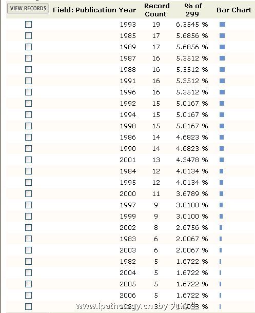
第一张,这片文章一共被引用了299次,这当然不包括中国学者的引用,主要是SCI引用,引用的高峰出现在1985-2002年,顶峰是1993年
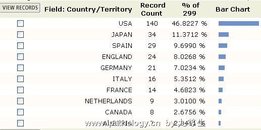
第二张 引用这片文章的最多国家是美国,几乎占了一半
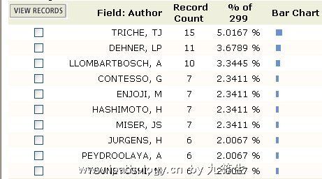
第三张 引用这片文章最多的头十位学者
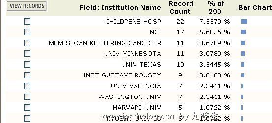
第四张 引用这片文章最多的10个机构
第五张 引用这片文章最多的领域,可以看出,绝大多数是病理方面的
第六张 引用这片文章最多的杂志
从第7至10张图片说明的是关于askin tumor方面的文章
第七张 发表askin tumor方面文章最多的作者,前十名
第八张 发表askin tumor方面文章最多的国家和地区,美国依然独占鳌头
第九张 研究askin tumor最多的机构
第十张 发表askin tumor的年代,不难看出,最为集中的年代是1992-1995,其次是1998-2001,而从2003年开始,这方面的文章开始减少(为什么)
- 达亦不足贵,穷亦不足悲
-
本帖最后由 于 2006-11-19 16:46:00 编辑
在分析完以上信息的时候,我发现一个有趣的东西,最早称呼askin tumor的并不是范教授引用的那两篇文献而是另有文献
LINNOILA RI, TSOKOS M, TRICHE TJ, et al.
EVIDENCE FOR NEURAL ORIGIN AND PERIODIC ACID SCHIFF-POSITIVE VARIANTS OF THE MALIGNANT SMALL CELL TUMOR OF THORACOPULMONARY REGION (ASKIN TUMOR)
LABORATORY INVESTIGATION 48 (1): A51-A51 1983
FINK IJ, KURTZ DW, CAZENAVE L, et al.
MALIGNANT THORACOPULMONARY SMALL-CELL (ASKIN) TUMOR
AMERICAN JOURNAL OF ROENTGENOLOGY 145 (3): 517-520 1985
时间上要更早一点:D
- 达亦不足贵,穷亦不足悲
由第10张图,我们不难发现,1992年是askin tumor研究的一个分水岭,这一年有什么进展让askin tumor的研究一下之热了一把?在此之前有些什么样的研究?
1992年以前的研究
Askin tumor具有神经分化(Gonzalezcrussi, Wolfson et al. 1984)和PAS阳性(Linnoila, Tsokos et al. 1983; Linnoila, Tsokos et al. 1986)。遗传学改变(Seemayer, Vekemans et al. 1985),推测可能来源于神经外胚层(Fujii, Hongo et al. 1989),最直接提及askin tumor是胸肺区pnet的文章(Faubert and Inniger 1991)
Faubert, C. and R. Inniger (1991). "Mri and Pathological Findings in 2 Cases of Askin Tumors." Neuroradiology 33(3): 277-281.
Askin tumors are primitive peripheral neuroectodermal tumors (PPNET) located in the thoracopulmonary region. This entity was first proposed in 1979 by Askin et al. These highly malignant tumors occur primarily in children and young adults. Pre- und postoperative MRI findings are presented for two pathologically proven cases. MRI is the most appropriate imaging modality for the diagnosis and eventual follow-up for these tumors. They appear homogenous, and iso- or discretely hypointense in comparison to the spinal cord on T1-weighted images and hyperintense on proton density and T2-weighted images. They show a very stark contrast enhancement after i.v. injection of paramagnetic contrast agents. Sharp tissue borders, and exact tumor extension were shown in both cases. The high signal intensity on T2-weighted images was not altered by chemotherapy.
Fujii, Y., T. Hongo, et al. (1989). "Cell-Culture of Small Round Cell Tumor Originating in the Thoracopulmonary Region - Evidence for Derivation from a Primitive Pluripotent Cell." Cancer 64(1): 43-51.
The authors describe a 14-year-old girl with small round cell tumor originating in the chest wall analyzed by the extensive studies including light and electron microscopic examination, histochemical study, immunochemical study, cytogenetics, and gene analysis. A cell line producing carcinoembryonic antigen (CEA) and neuron-specific enolase (NSE) has been established from pleural effusion of the pulmonary metastatic tumor. Cytogenetic analysis disclosed a reciprocal translocation (11;22)(q24;q12). Additionally, immunocytochemical studies demonstrated that CEA, NSE, vimentin, cytokeratin, and epithelial membrane antigens are positive, but desmin and S-100 protein are negative. Although neurofilament was negative in the pulmonary metastatic tumor cells, it became positive in cell line in vitro. These results suggest that this tumor may be derived from the primitive and pluripotential cells, differentiating into mesenchymal, epithelial, and neural features in variable proportions.
Gonzalezcrussi, F., S. L. Wolfson, et al. (1984). "Peripheral Neuroectodermal Tumors of the Chest Wall in Childhood." Cancer 54(11): 2519-2527.
A retrospective review of primary chest wall malignant tumors of childhood collected at the Children's Memorial Hospital of Chicago was undertaken. Among twelve instances of poorly differentiated neoplasms whose uniform, monotonous structure made accurate classification difficult or impossible by conventional histologic study, there were three tumors with features suggestive of neuroectodermal differentiation. Electron microscopic and immunohistologic findings further strengthened this interpretation, despite the fact that none of the patients had evidence of a primary neuroblastoma outside the chest wall. These results and a review of the pertinent literature support the conclusion that neuroectodermal neoplasms in childhood may present in peripheral somatic tissues with greater frequency than is commonly assumed. The importance of this distinction is discussed, particularly the need to distinguish these neoplasms from Ewing's sarcoma.
Linnoila, R. I., M. Tsokos, et al. (1983). "Evidence for Neural Origin and Periodic Acid Schiff-Positive Variants of the Malignant Small Cell Tumor of Thoracopulmonary Region (Askin Tumor)." Laboratory Investigation 48(1): A51-A51.
Linnoila, R. I., M. Tsokos, et al. (1986). "Evidence for Neural Origin and Pas-Positive Variants of the Malignant Small-Cell Tumor of Thoracopulmonary Region (Askin Tumor)." American Journal of Surgical Pathology 10(2): 124-133.
The differential diagnoses of childhood and adolescent tumors composed of small round cells include a distinctive clinicopathological entity called malignant small cell tumor (MSCT) of the thoracopulmonary region in childhood. In the present study, 15 such tumors that fulfilled the criteria by Askin et al. were examined for features of possible neural differentiation by light and electron microscopy (EM). With hematoxylin-eosin stain (H&E) the tumors were made up of small undifferentiated cells; rosette formation was noticed in four cases. By immunohistochemistry all 15 tumors were positive for neuron/specific enolase (NSE), which is a specific marker for neural elements and their tumors including neuroblastomas. Ten of 15 MSCT had positive PAS staining. Ultrastructurally dense core (neurosecretory) granules and cell processes indicative of neuronal differentiation could be recognized in 10 of 14 tumors. The dense core granules were often atypical. Filamentous cytoskeleton, never observed in Ewing's sarcoma, was often present. Based on the current results, MSCT of the thoracopulmonary region can be considered a peripheral neuroectodermal tumor with the possible origin in intercostal nerves. MSCTs are generally misdiagnosed as Ewing's sarcoma due to their primitive appearance in H&E sections and their periodic acid-Schiff positivity. NSE immunostaining, preferably augmented by electron microscopy, is necessary for their correct diagnosis.
Seemayer, T. A., M. Vekemans, et al. (1985). "Histological and Cytogenetic Findings in a Malignant-Tumor of the Chest-Wall and Lung (Askin Tumor)." Virchows Archiv a-Pathological Anatomy and Histopathology 408(2-3): 289-296.
This report describes a histological and cytogenetic study of a malignant tumor involving the chest wall and lung (Askin tumor) of a young girl. Although initially considered to represent a variant of Ewing's sarcoma, immunocytochemical studies disclosed neuron-specific enolase in neoplastic cells. Ultrastructural study revealed rare cells which contained microtubules and/or dense core neurosecretory type granules. Cytogenetic analysis of neoplastic cells disclosed a reciprocal translocation (11;22)(q24;q12) and occasional extrachromosomal structures interpreted as double minute chromosomes. The latter finding, an indication of gene amplification, is commonly identified in neural crest-derived neoplasms. These ultrastructural, immunocytochemical, and karyotypic data provide evidence in support of a neuroepithelial histogenesis for the Askin tumor.
- 达亦不足贵,穷亦不足悲
-
本帖最后由 于 2006-11-19 19:31:00 编辑
1992年发生了什么?
我们需要看看范教授的第四篇参考文献
Contesso, G., A. Llombartbosch, et al. (1992). "Does Malignant Small Round Cell Tumor of the Thoracopulmonary Region (Askin Tumor) Constitute a Clinicopathological Entity - an Analysis of 30 Cases with Immunohistochemical and Electron-Microscopic Support Treated at the Institute-Gustave-Roussy." Cancer 69(4): 1012-1020.
The morphology and clinical outcome of 30 patients with malignant small round cell tumors located in the thoracopulmonary region (Askin tumor) are reported. Histologically, all tumors had similar patterns, with small round-to-oval cells and a lobulated stroma. Immunohistochemical analysis always resulted in positive staining for one or several neural markers. No significant differences were found compared with the immunomarkers in 26 typical Ewing's sarcomas located outside the thoracic wall. In three specimens, electron microscopy confirmed the presence of membrane-bound neurosecretory granules. It was confirmed that there is a remarkable similarity among all malignant small round cell tumors, including Askin tumor and Ewing's sarcoma. Overall survival was poor with a 2-year rate of 38% and a 6-year rate of 14%.
Dehner, L. P. (1993). "Primitive Neuroectodermal Tumor and Ewings-Sarcoma." American Journal of Surgical Pathology 17(1): 1-13.
Many of the major solid, malignant tumors of childhood have histologic similarities that reflect their dysembryonic and primitive features. One subset of these neoplasms, Ewing's sarcoma (ES) and primitive neuroectodermal tumor (PNET), presents primarily in the bone and soft tissues. Both tumor types were reported at a time and date well before the advent of electron microscopy and immunohistochemistry. Opposition to ES and PNET as distinctive entities developed and persisted because these tumors were considered incompletely documented examples of metastatic neuroblastoma or malignant lymphoma. General acceptance of ES as a unique tumor type occurred well before the PNET had been fully defined and characterized. Once these neoplasms had joined the other round cell neoplasms, the quest for the histogenesis was pursued, but the results were frustratingly inconclusive, especially for ES. Because of the resemblance of the PNET to classic neuroblastoma, the neural crest was regarded as the most likely progenitor. With the recognition of osseous PNET, extraosseous ES, and a shared cytogenetic abnormality between ES and PNET, more recent speculation has focused on the possibility that these presumably separate neoplasms are closely related histogenetically without directly answering the question of histogenesis. Despite the likely common progenitorship of ES and PNET, the latter neoplasm is seemingly the more aggressive. Although melanotic neuroectodermal tumor of infancy, intra-abdominal desmoplastic small cell tumor, and polyphenotypic small cell tumors have some overlapping microscopic and immunohistochemical features with PNET, their relationship to ES-PNET has otherwise not been resolved.
Dierick, A. M., H. Roels, et al. (1993). "The Immunophenotype of Ewings-Sarcoma - an Immunohistochemical Analysis." Pathology Research and Practice 189(1): 26-32.
Since 1963, 53 cases of Ewing's sarcoma have been diagnosed at the N. Goormaghtigh Institute of Pathological Anatomy. All tumours were classified according to their growth pattern as described by Kissane: diffuse, lobular or organoid, the latter with pseudorosettes, or belonging to the fillagree type. In 45 cases, an immunohistochemical examination was carried out on routinely processed material in order to determine the immunophenotype of the tumours and to get more information on the histogenesis of the tumours. Antibodies against cytokeratin, desmin, vimentin, neurofilaments, neuron-specific enolase, Leu7, synaptophysin, chromogranin, the muscle-specific actin, the Von Willebrand factor and the common leucocyte antigen were used in a biotin-streptavidin procedure. The presence of neural markers such as neuron-specific enolase, synaptophysin and Leu7 was proved in the majority of the cases. The expression of the three markers was absent in only two cases. Neurofilaments were demonstrated to be present in S of the 6 cases for which fresh material was available. The results of this study point towards a possible neuroectodermal origin of Ewing's sarcoma. This is in agreement with the results of cell culture experiments and of cytogenetic studies.
Shishikura, A., S. Ushigome, et al. (1993). "Primitive Neuroectodermal Tumors of Bone and Soft-Tissue - Histological Subclassification and Clinicopathological Correlations." Acta Pathologica Japonica 43(4): 176-186.
Recent reports of Ewing's sarcoma (EW) and extraskeletal Ewing's sarcoma (EEW) support the hypothesis that these tumors are neuroectodermal in origin. Primitive neuroectodermal tumors (PNET) of bone (32 cases) and soft tissue (25 cases) including those previously categorized as EW in 27 cases and EEW in 15 cases were carefully studied histologically, immunocytochemically and morphometrically, focusing on tumor cell differentiation. This study attempts to subclassify these tumors on the basis of the size of tumor cells and nuclei, their variations (uniformity or diversity), arrangement of tumor cells (rosette or non-rosette), focal differentiation to larger ganglion-like cells, and staining intensity for neural markers. All tumors were histologically subclassified as small, medium or large cell types, three basic subtypes (rosette type, abortive rosette type, non-rosette type) and four complementary subtypes (fibrillary type, non-fibrillary type, angiomatoid type, ganglion cell type). Classic EW or EEW is consistent with small or medium, non-rosette, non-fibrillary type tumors, previously described large cell EW with large, non-rosette, fibrillary or non-fibrillary type tumors, and classic neuroectodermal tumor with small or medium, rosette, fibrillary type tumors, according to the present subclassification. Clinicopathologic correlations with the different subtypes are discussed. Long-term survival, more than 5 years, was seen in patients with small cell type, and those younger than 14 years of age.- 达亦不足贵,穷亦不足悲
努力的试图区分PNET和骨外EWS
Brinkhuis, M., L. C. D. Wijnaendts, et al. (1995). "Peripheral Primitive Neuroectodermal Tumor and Extra-Osseous Ewings-Sarcoma - a Histological, Immunohistochemical and DNA Flow Cytometric Study." Virchows Archiv-an International Journal of Pathology 425(6): 611-616.
Although peripheral primitive neuroectodermal tumour (pPNET) and extra-osseous Ewing's sarcoma (EES) are thought to be closely related neoplasms, their clinical behaviour differs considerably. To determine the clinical relevance of the Schmidt classification scheme for differentiating pPNET and EES, 20 tumour specimens of poorly differentiated round cell tumours were evaluated. In addition, the diagnostic value of several neural markers and the prognostic value of quantitative morphological variables (DNA ploidy, S-phase fraction, and the mitotic activity) were assessed. Homer-Wright rosettes were present in 9 tumours. Neuron specific enolase (NSE) was expressed in 11 tumours, 8 of which expressed a second neural marker (CD57, S100, or neurofilament). According to the Schmidt classification, 11 pPNET and 5 EES were distinguished. HBA-71 was exclusively expressed in pPNET and EES. The remaining tumours were classified as sarcoma not otherwise specified (n = 2), rhabdomyosarcoma (n = 1), and desmoplastic tumour with divergent differentiation (n = 1). EES611 patients fared significantly better than the pPNET patients (100% versus 42% 5-year survival). Neither DNA ploidy nor S-phase fraction assessed in 12 evaluative histograms (9 pPNET and 3 EES), nor mitotic activity yielded information of additional prognostic value. On the basis of this study and the Schmidt classification scheme, it can be concluded that if the diagnosis of EES and pPNET is based on light microscopy (Homer-Wright rosettes) and/or immunohistochemistry (at least two neural markers, i.e. NSE, S-100, CD57, and neurofilament), the classification provides important clinical information. Furthermore, positivity for HBA-71 is helpful in differentiating pPNET and EES from all other small round cell tumours.
- 达亦不足贵,穷亦不足悲
1995年的归纳
Vonschlippe, M. and J. S. Whelan (1995). "Primitive Neuroectodermal Tumor of the Chest-Wall." Annals of Oncology 6(4): 395-401.
The concept of primitive neuroectodermal tumour (PNET) has been evolving for many years, as has its nomenclature. It was first described as a tumour arising in peripheral nerve, when it was called neuroepithelioma [1]. These tumours are part of the differential diagnosis of malignant small round cell tumours, which include Ewing's sarcoma, rhabdomyosarcoma, neuroblastoma and lymphoma, and which appear as sheets of monotonous small round cells on light microscopy, staining dark blue with haematoxylin and eosin. In the 1970's, reports of tumours displaying neural features introduced new terms such as primitive neuroectodermal tumour and peripheral neuroectodermal tumour. A relationship to the undifferentiated primitive neuroectodermal tumours of the central nervous system in children was assumed by the title of these tumours, although it was recognised that they were not necessarily related to peripheral nerves. In 1979, Askin described a series of patients with malignant small cell tumours of the chest wall which showed neural differentiation, but which bore a resemblance to Ewing's tumour [2]. Later, this tumour was also diagnosed at other sites, and became identified with the previously described neuroepitheliomas, but the names multiplied, e.g. peripheral neuroepithelioma, peripheral neuroblastoma, Askin tumour, and peripheral PNET. The term 'primitive neuroectodermal tumour' is used in this article. Cytogenetic studies have confirmed that it forms part of a spectrum with both classical and atypical Ewing's tumour [3]. The clinical and pathological features of PNET, its management, and perspectives for the future, are discussed with reference to a case of PNET of the chest wall.
到此是不是有一点清楚了?
- 达亦不足贵,穷亦不足悲
我们又看到一个新的称呼Ewing's sarcoma family(Adams, Hany et al. 1996)
Adams, V., M. A. Hany, et al. (1996). "Detection of t(11;22)(q24;q12) translocation breakpoint in paraffin-embedded tissue of the Ewing's sarcoma family by nested reverse transcription-polymerase chain reaction." Diagnostic Molecular Pathology 5(2): 107-113.
Tumors of the Ewing's sarcoma family often present a major diagnostic challenge for the pathologist. In recent years, significant progress has been made in identifying characteristic chromosomal rearrangements associated with certain solid tumors. More than 85% of Ewing's sarcoma and related tumors present a specific t(11;22) (q24;q12) balanced translocation, which generates a fusion transcript of the EWS gene and the FLI-1 gene. The cloning of the t(11;22)(q24;q12) breakpoint has raised the possibility of using a reverse transcription-polymerase chain reaction (RT-PCR) based assay as a diagnostic tool. We report an improvement of the established method, which currently depends on fresh or snap-frozen tissue, so that it is possible to use formalin-fixed, paraffin-embedded tissue as a source of RNA. The described nested RT-PCR assay enables the pathologist to investigate retrospectively archival tumor samples or to confirm the diagnosis in cases where no fresh or frozen material is available.
- 达亦不足贵,穷亦不足悲
2000年的综述(de Alava and Gerald 2000)
de Alava, E. and W. L. Gerald (2000). "Molecular biology of the Ewing's sarcoma/primitive neuroectodermal tumor family." Journal of Clinical Oncology 18(1): 204-213.
Ewing's sarcoma (ES) and primitive neuroectodermal tumor (PNET) are members of a tumor family consistently associated with chromosomal translocation and functional fusion of the EWS gene to any of several structurally related transcription factor genes. Similar gene fusion events occur in other mesenchymal and hematopoietic tumors and are tumor-specific. The resulting novel transcription factor-like chimeric proteins are believed to contribute to tumor biology by aberrant regulation of gene expression altering critical controls of cell proliferation and differentiation. These tumor-specific molecular rearrangements are useful for primary diagnosis, may provide prognostic information, and present potential therapeutic targets. The recent advances in our understanding of the molecular biology of ES and PNET represent a paradigm for the application of the basic biology of neoplasia to clinical management of patients. (C) 2000 by American Society of Clinical Oncology.2001年的综述(de Alava and Pardo 2001)
de Alava, E. and J. Pardo (2001). "Ewing tumor: Tumor biology and clinical applications." International Journal of Surgical Pathology 9(1): 7-17.
The Ewing tumor family includes classical Ewing's sarcoma of bone and soft tissues, peripheral primitive neuroectodermal tumors (pPNET), Askin tumor, and other less frequent variants. This group of tumors is defined hy the consistent presence of chromosomal translocations resulting in gene fusions between EWS gene and a member of the ETS family of transcription factors, mainly FLI1 and ERG. Analogous fusions are seen in other solid developmental tumors, like desmoplastic small round cell tumor. These fusions, which are consistently present and tumor-specific, control transcription of several target genes, largely unknown but critical to cell proliferation and differentiation. Therefore. gene fusions are useful to diagnose and classify small round cell tumors, have prognostic significance, are probably useful to detect micrometastasis and monitor minimal residual disease, and are potential therapeutic targets. Secondary molecular alterations, which include mutations of cell cycle regulatory genes, are not tumor-specific but are related to progression and may have prognostic value. The Ewing rumor family represents a paradigm of the application of the knowledge of biology of neoplasia to the clinical management of patients.
- 达亦不足贵,穷亦不足悲
2001年who,将三者合并,变成了EWS/pnet。具体内容参阅WHO(2001)分类
Soft tissue tumor(第三版)也作了同样的归类。好像也是2001年出版的
2003年的研究(Burchill 2003)
Burchill, S. A. (2003). "Ewing's sarcoma: diagnostic, and therapeutic implications of molecular abnormalities." Journal of Clinical Pathology 56(2): 96-102.
The identification of the non-random chromosome rearrangements between the EWS gene on chromosome 22q12 and members of the ETS gene family in Ewing's sarcoma, peripheral primitive neuroectodermal tumour, Askin tumour, and neuroepithelioma has been a key advance in understanding their common histogenesis and defining the Ewing's sarcoma family of tumours (ESFT). In addition to improvements in diagnosis and potentially the stratification of patients for risk, biological investigations of these gene fusions may define targets for much needed therapeutic strategies to eliminate minimal residual disease or metastatic disease. Insight into their relation with other oncogenic events in ESFT will advance risk group analysis and ultimately may improve clinical management and survival for patients with this disease.
Yoshino, N., T. Kojima, et al. (2003). "Diagnostic significance and clinical applications of chimeric genes in Ewing's sarcoma." Biological & Pharmaceutical Bulletin 26(5): 585-588.
Ewing's sarcoma (ES) is one of the most malignant bone and soft tissue tumors in childhood. Morphologically, ES belongs to the small round cell tumors (SRCT). ES, peripheral primitive neuroectodermal tumor (PNET), and Askin's tumor are classified as ES family tumors (ESFT) because they share a common chromosomal translocation. The EWS-FLI1 chimeric gene is generated by t (11; 22). Other reciprocal translocations resulting in formation of chimeric genes between EWS and ETS family genes (ERG, ETV1, E1AF, and FEV) are t (21; 22), t (7; 22), t (17; 22), and t (2; 22), respectively. Although it is generally difficult to distinguish ES from SRCT, we could easily and quickly distinguish ES from other SRCT by using reverse transcription polymerase chain reaction (RTPCR). We looked for specific chimeric genes in 23 tumor samples, including three ES clinical samples. We detected five chimeric genes in the three ES samples. Three chimeric genes, all EWS-FLI1, were detected in one ES sample. Different chimeric genes, EWS-ERG and EWS-ETV1, were detected in the other two ES samples. Moreover, because we could not detect specific chimeric genes in samples from non-ESFT, it may be possible to use this technique to diagnose ESFT and to detect tumor cell contamination before hematopoietic stem cell transplantation.
- 达亦不足贵,穷亦不足悲


