| 图片: | |
|---|---|
| 名称: | |
| 描述: | |
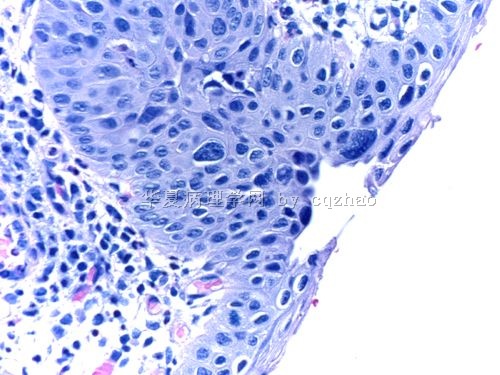
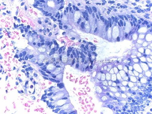
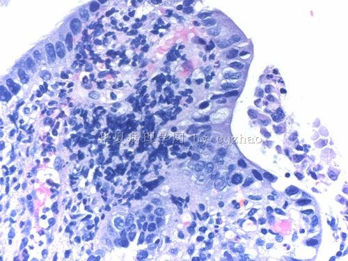
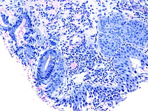
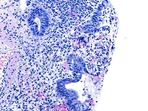
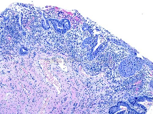
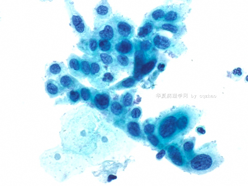
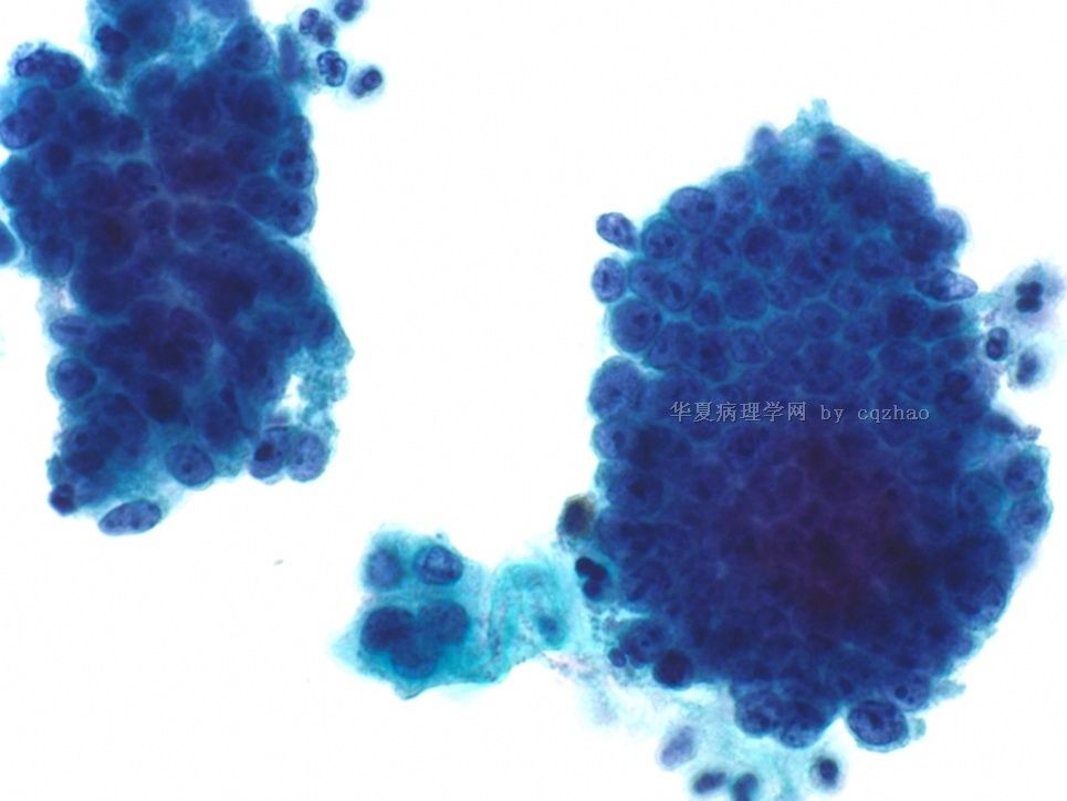
- 25 y/f Pap test (cqz c-19)
| 以下是引用viivi薇在2011-2-16 22:32:00的发言: 请问赵老师,HSIL累腺与AGC合并HSIL有什么样的鉴别要点?组织学也是和ABIN老师的一样,CIN2累腺 |
HSIL累腺与AGC合并HSIL有什么样的鉴别要点?It is very difficult in cytology.
HSIL累腺: Some times flattening of cells is present at the edge of the cluster of high grade cells.
AIS: Feathering, palisading et al.
In practice it will be difficult.
| 以下是引用有福不在忙在2011-2-16 10:26:00的发言: 染P16和KI67看看吧,鳞化是比较典型的,看看是否伴有CIN。 |
在中国做医生你真是有福啊。
You may have different feeling when you read the glass slides and photos.
This should be a case to be signed out with 几分钟reading and thinking.
I signed out the case already. Do you think I need to do the stains?
-
本帖最后由 于 2011-02-13 03:14:00 编辑
I have not read biopsy slides yet. In other words I do not know the histological result now. Definitely I will have biopsy slides and paste photos here in Monday. You guys can continue to discuss the case. It is a good opportunity to show your bright part (kiding). In fact the interpretation based on a few photos on line and true glass slides can be much different. We come here for study. No persons care you make wong interpretation.
Remember that the cases of Pap cytology with histologcal follow up result are best ones for study.
| 以下是引用巴山夜雨涨秋池在2011-2-12 19:20:00的发言: 分歧很大。当这么多人都把第一图看成ASCH或更低时,我就开始怀疑我的显示屏或眼睛是否出了问题。尽管分歧如此大,我仍要表达个人意见:图一应当是HSIL。无论倍数大小,关键在找参照。图一的参照细胞在哪里呢?我认为图一是一堆核增得很大的细胞,且有染色质异常及深染,核畸形是明显的,不用怀疑其为高级别的。图二马兄的分析与我不谋而合。欢迎继续争论,并期待赵老师指导。 |
