| 图片: | |
|---|---|
| 名称: | |
| 描述: | |
- 小脑肿瘤,会诊!!
-
cnlzh20060 离线
- 帖子:224
- 粉蓝豆:58
- 经验:378
- 注册时间:2009-02-27
- 加关注 | 发消息
| 姓 名: | ××× | 性别: | 男 | 年龄: | 56 |
| 标本名称: | 小脑肿瘤 | ||||
| 简要病史: | 肺部有一占位,未手术。 | ||||
| 肉眼检查: | 灰红组织,4*3*3cm | ||||
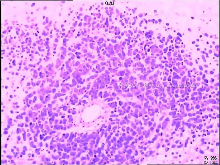
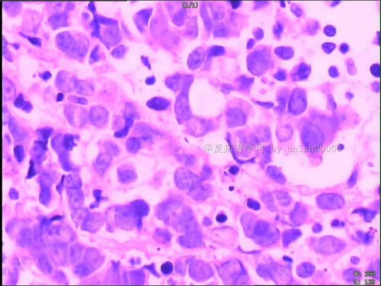
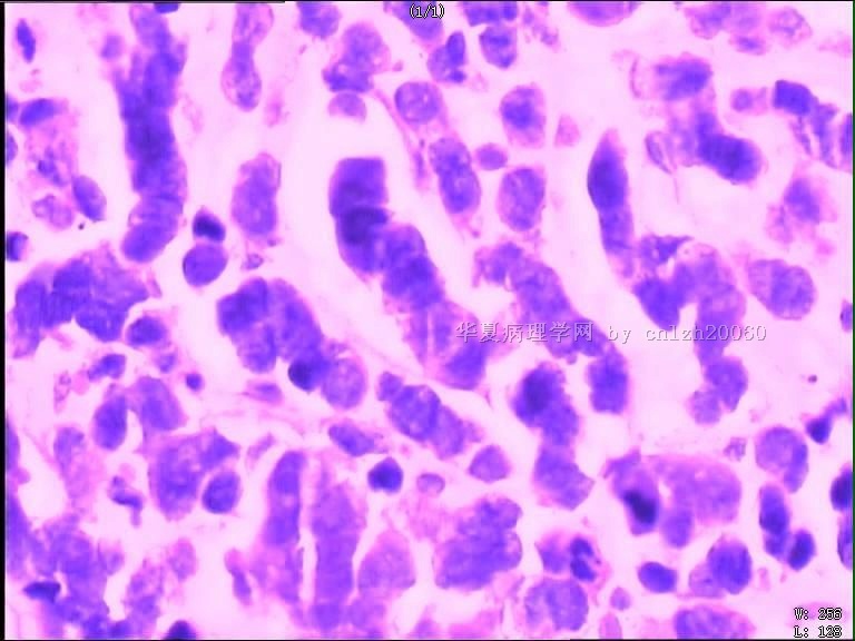
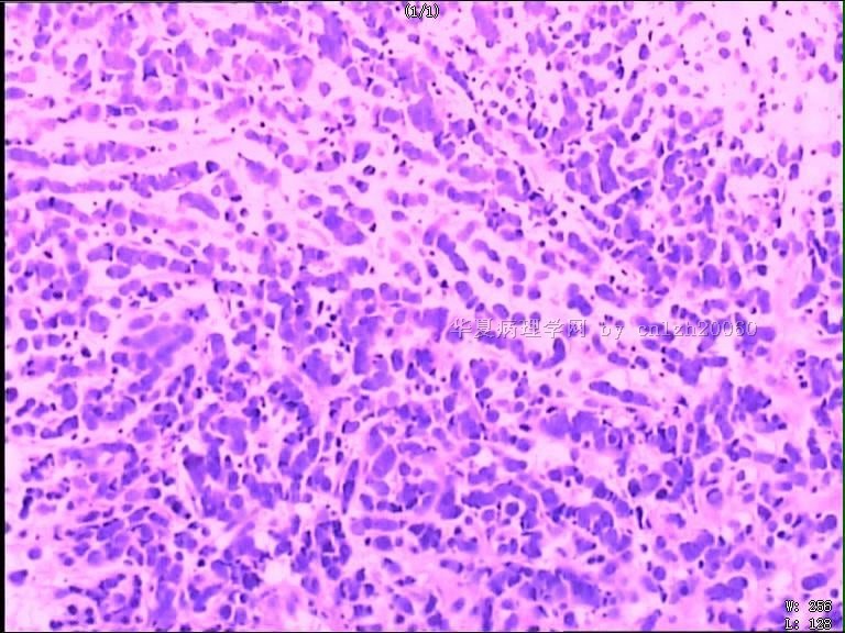
镜下见:局灶坏死,核分裂像易见约10个/HP,细胞排列成流线样。已做免疫组化,结果为Syn(+),CK局灶(+),GFAP(-),TTF-1(-),CD99(+)。
疑问:
1.如考虑为不典型类癌,应是肺源性可能性大,未见器官样结构,TTF-1(-);
2.如考虑为髓母细胞瘤,患者年龄偏大;CK局灶(+),这一点少见(阿克曼上写个别病例可以CK局灶+);
3.如考虑小细胞癌,细胞形态又不像。
名称:图1
描述:图1
名称:图2
描述:图2
名称:图3
描述:图3
名称:图4
描述:图4
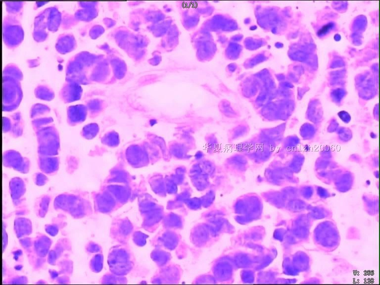
名称:图5
描述:图5
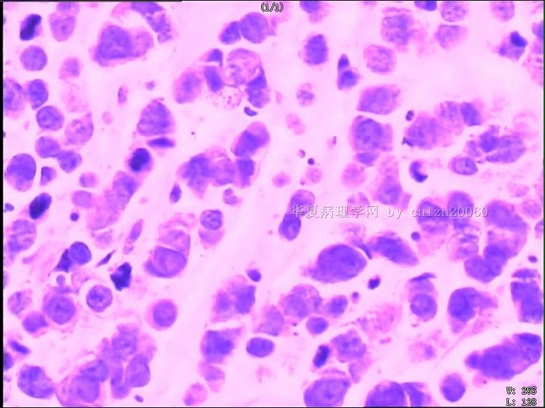
名称:图6
描述:图6
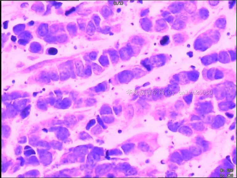
名称:图7
描述:图7
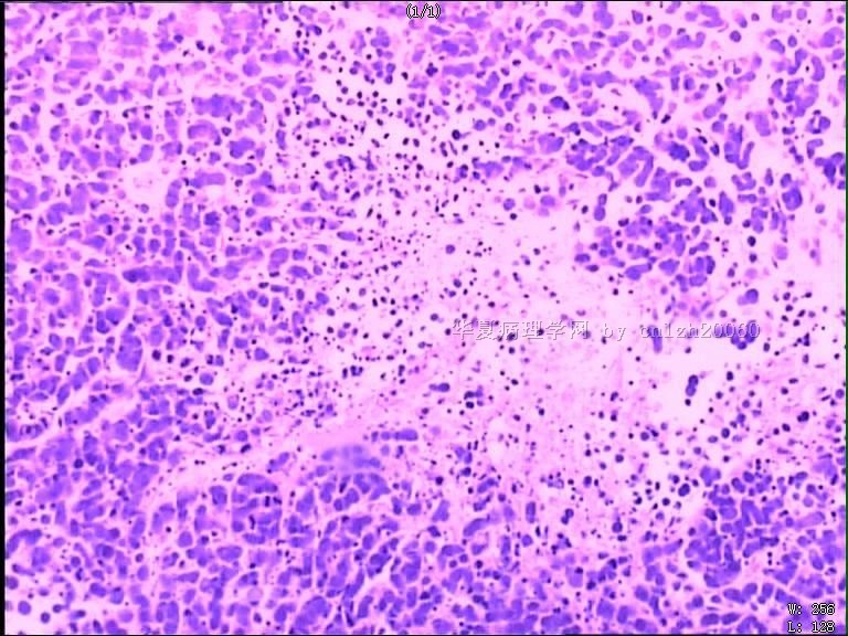
名称:图8
描述:图8
标签:
×参考诊断
-
As said already, this is not a case of atypical carcinoid tumor with so many mitoses and focal tumor necrosis seen. If cytokeratins stain is indeed positive and GFAP stain is indeed negative, this is unlikekly medulloblastoma of cerebellum and likely metastatic small cell carcinoma from lung.

聞道有先後,術業有專攻
| 以下是引用mjma在2010-10-25 20:56:00的发言: As said already, this is not a case of atypical carcinoid tumor with so many mitoses and focal tumor necrosis seen. If cytokeratins stain is indeed positive and GFAP stain is indeed negative, this is unlikekly medulloblastoma of cerebellum and likely metastatic small cell carcinoma from lung. |


