


| 图片: | |
|---|---|
| 名称: | |
| 描述: | |
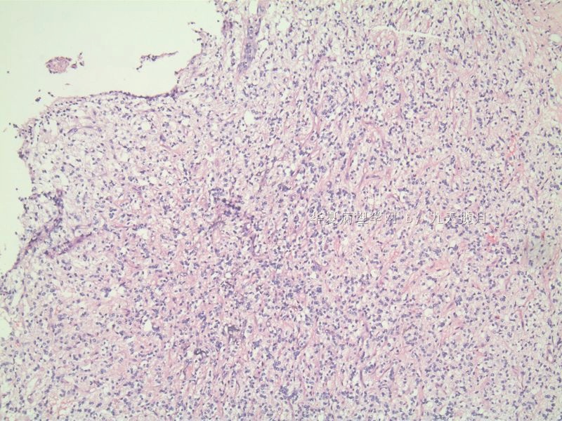
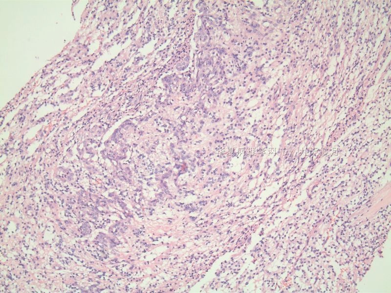
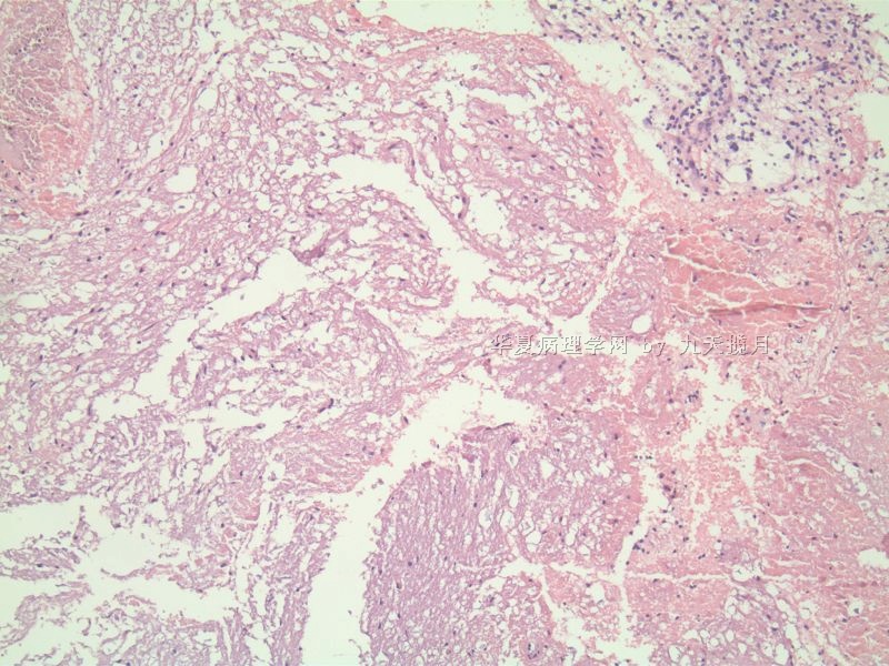
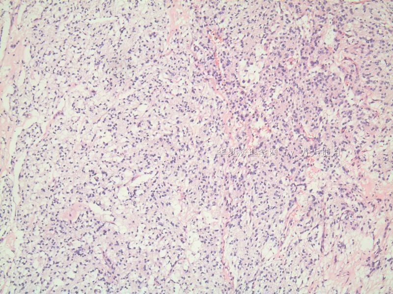
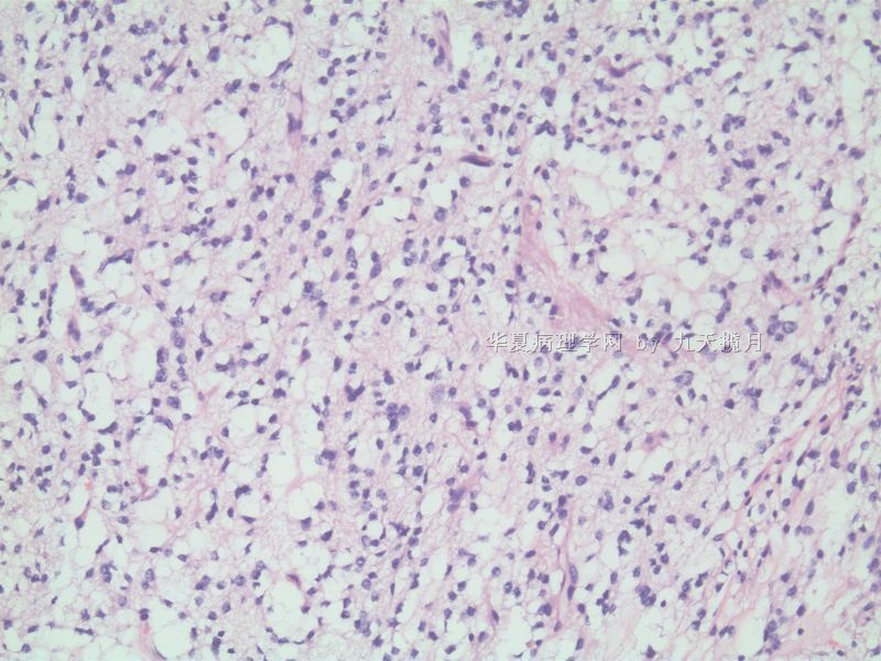
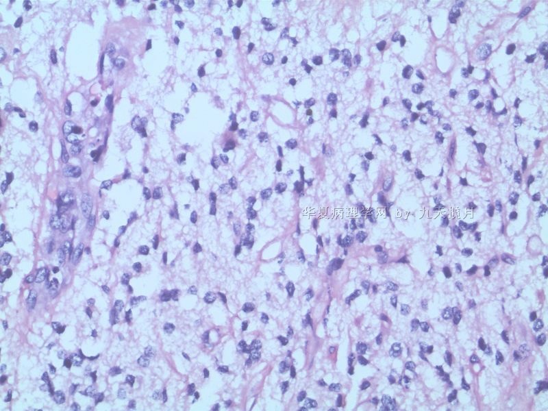
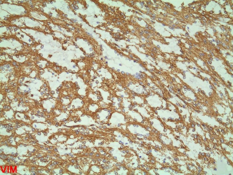
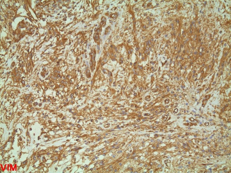
- 冰冻 9岁男,基底节占位
-
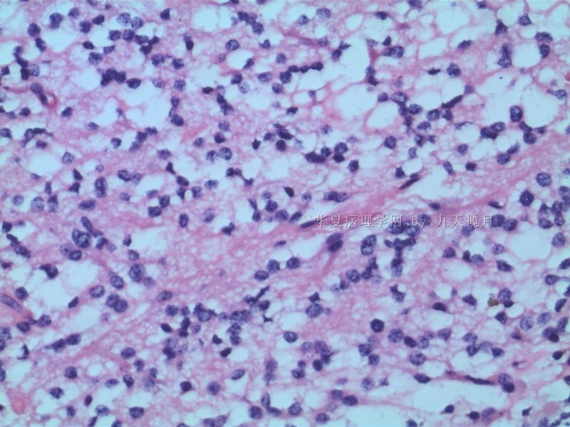
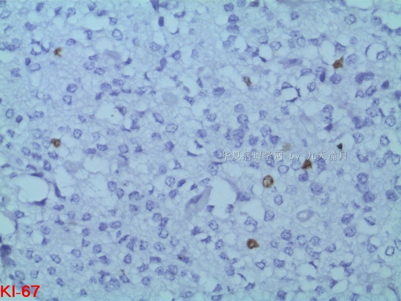
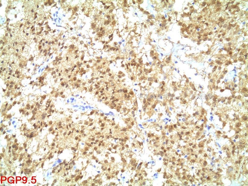
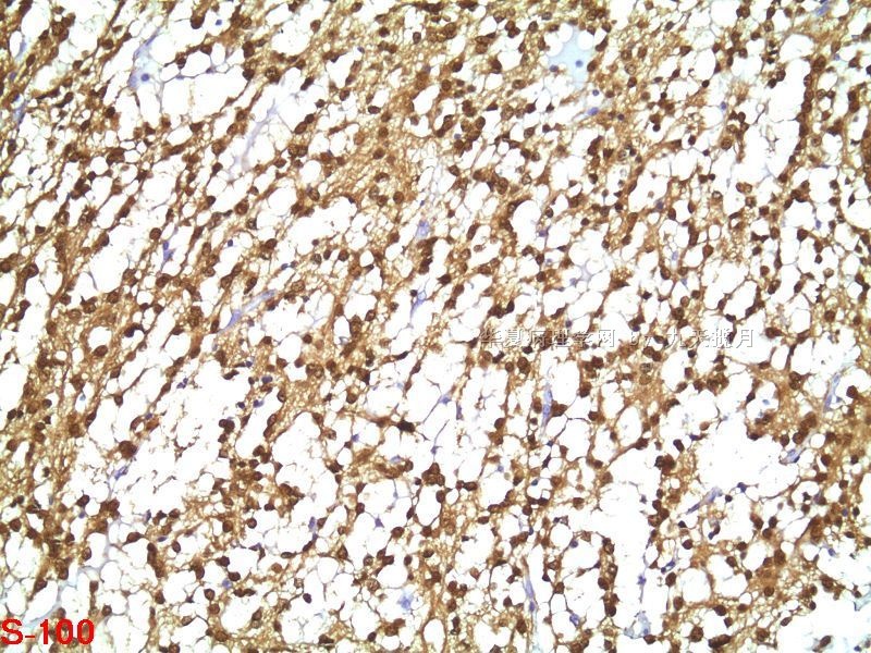
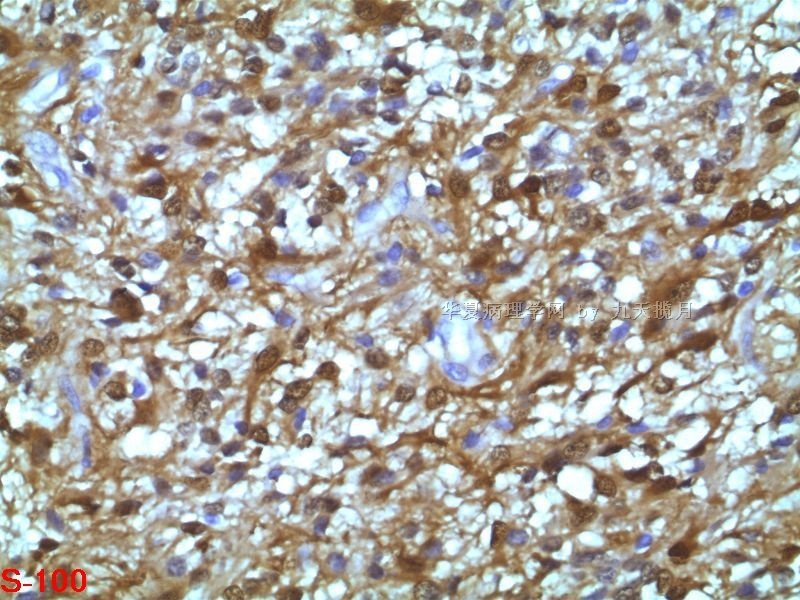
This appears to be a small stereotactic biopsy of a basal ganglionic tumor found incidentally. The boy has very mild neurologic deficit. Other than an infiltrative malignancy, it is very difficult to give further classification and grade of this tumor. In the absence of an epithelial element, I do not favor a mixed germ cell tumor. In the absence of a spindled cell mesenchymal component and large rhabdoid cells, I do not favor atypical rhabdoid/teratoid tumor (AT/RT). The degree of nuclear pleomorphism is relatively high, and I think this may be an anaplastic glioma (such as ependymoma) or a supratentorial PNET. I hope there was enough tissue not frozen for permanent sections.

聞道有先後,術業有專攻
| 以下是引用mjma在2010-8-24 7:55:00的发言: This appears to be a small stereotactic biopsy of a basal ganglionic tumor found incidentally. The boy has very mild neurologic deficit. Other than an infiltrative malignancy, it is very difficult to give further classification and grade of this tumor. In the absence of an epithelial element, I do not favor a mixed germ cell tumor. In the absence of a spindled cell mesenchymal component and large rhabdoid cells, I do not favor atypical rhabdoid/teratoid tumor (AT/RT). The degree of nuclear pleomorphism is relatively high, and I think this may be an anaplastic glioma (such as ependymoma) or a supratentorial PNET. I hope there was enough tissue not frozen for permanent sections. |
-
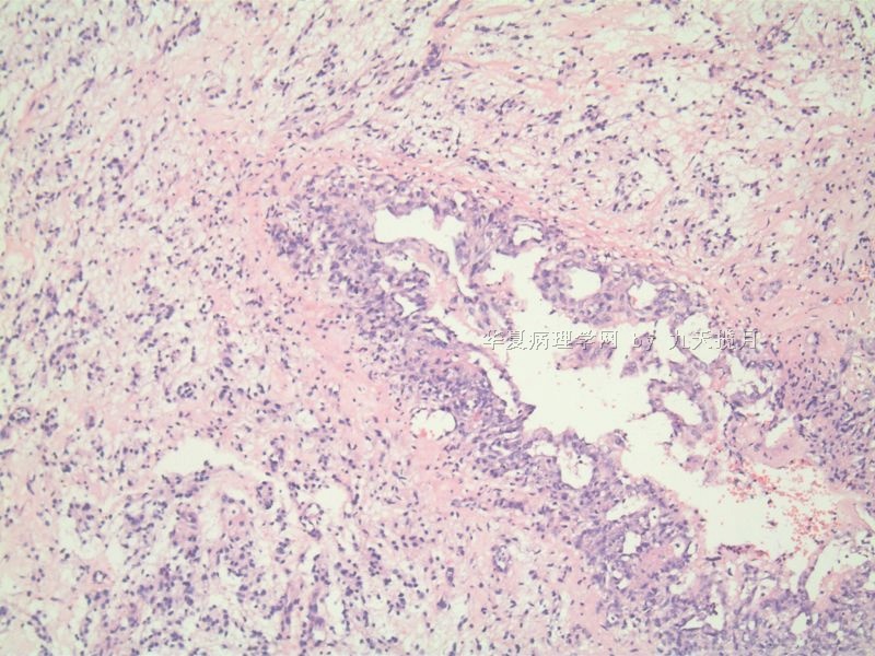
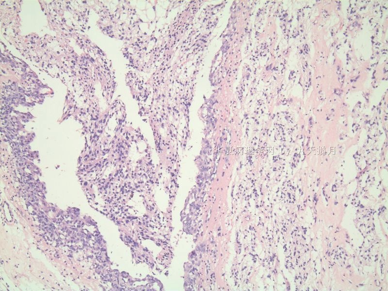
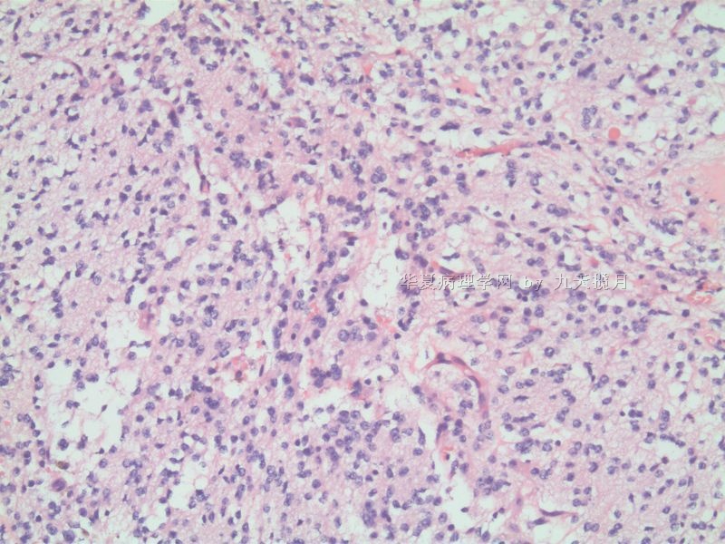
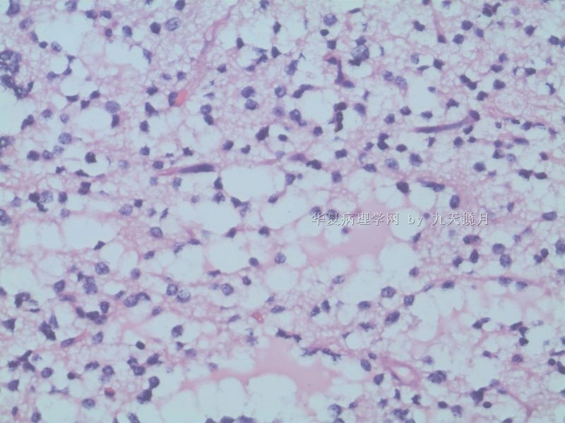
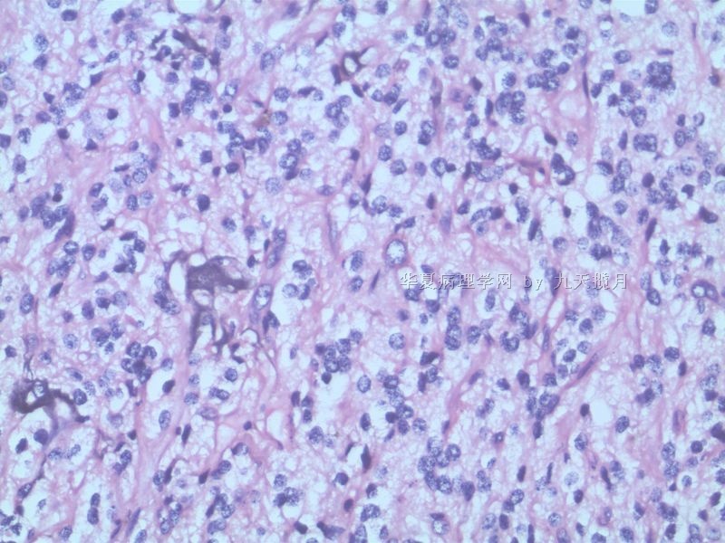
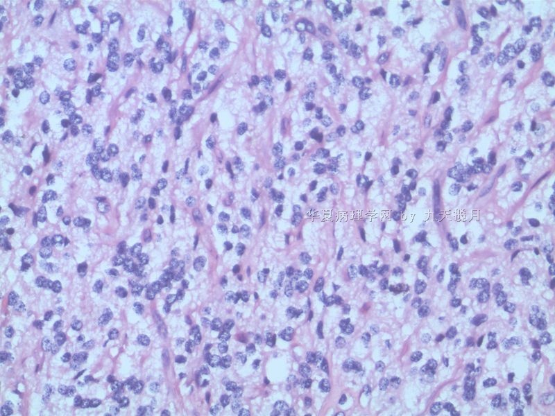
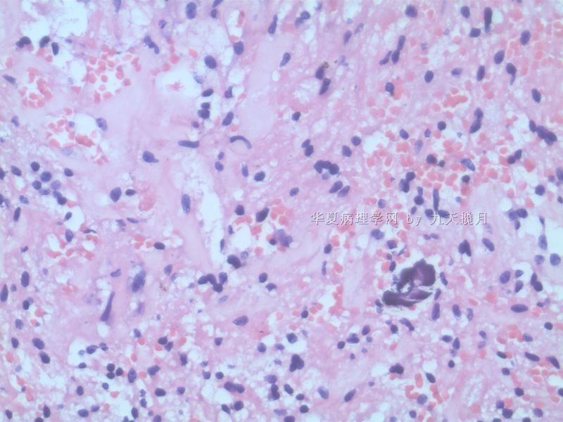
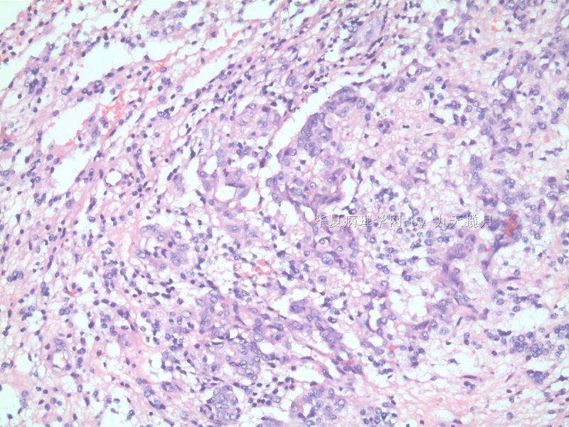
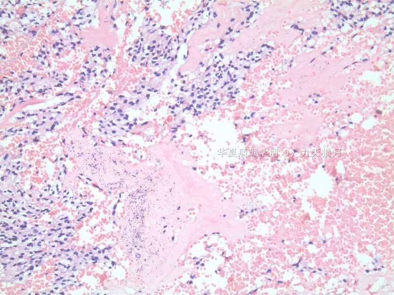
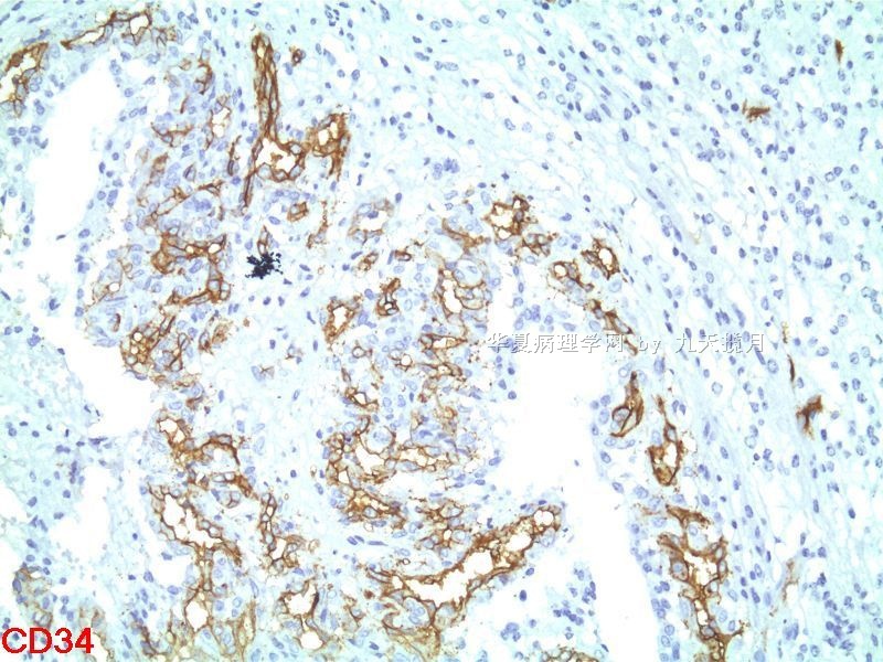
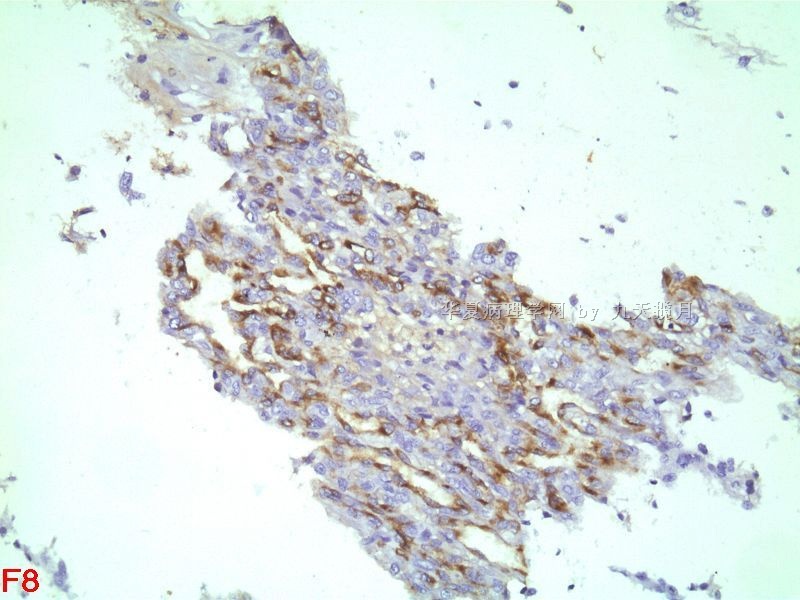
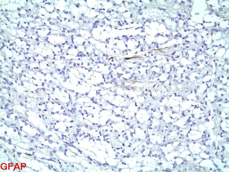
From the abundant tissue submitted for permanent sections, I must have been mistaken and the surgery was probably a resection and not a stereotactic biopsy. This is clearly a glioma. There are garlands of new vessels consistent with vascular/endothelial proliferation, but this may be the vascular lining of cystic areas in the tumor. Overall the tumor cells are fairly uniform in nuclear size and shape. There are calcospherites with or without associated small blood vessels. I do not find mitosis, severe nuclear pleomorphism/atypia, or tumor necrosis. Can you verify that mitotic figures are very rare to find in this case? If so, please look carefully for eosinophilic granular bodies and other features of pilocytic astrocytoma. I do not see dysplastic neurons or features of ependymal differentiation. Reviewing pre-surgical MRI would help confirm that this was a partially cystic, partially solid, circumscribed and enhancing lesion that may turn out to be WHO grade I pilocytic astrocytoma, which often mimic oligodendroglioma and fibrillary astrocytoma.
聞道有先後,術業有專攻
| 以下是引用mjma在2010-8-24 22:13:00的发言: From the abundant tissue submitted for permanent sections, I must have been mistaken and the surgery was probably a resection and not a stereotactic biopsy. This is clearly a glioma. There are garlands of new vessels consistent with vascular/endothelial proliferation, but this may be the vascular lining of cystic areas in the tumor. Overall the tumor cells are fairly uniform in nuclear size and shape. There are calcospherites with or without associated small blood vessels. I do not find mitosis, severe nuclear pleomorphism/atypia, or tumor necrosis. Can you verify that mitotic figures are very rare to find in this case? If so, please look carefully for eosinophilic granular bodies and other features of pilocytic astrocytoma. I do not see dysplastic neurons or features of ependymal differentiation. Reviewing pre-surgical MRI would help confirm that this was a partially cystic, partially solid, circumscribed and enhancing lesion that may turn out to be WHO grade I pilocytic astrocytoma, which often mimic oligodendroglioma and fibrillary astrocytoma. |


