共2页/29条首页上一页12下一页尾页
回复:63 阅读:10517
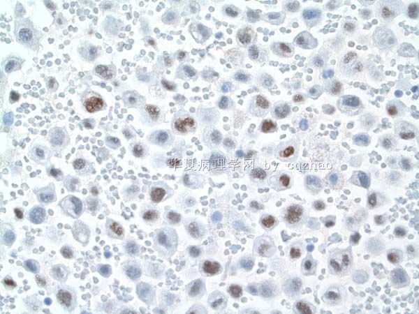
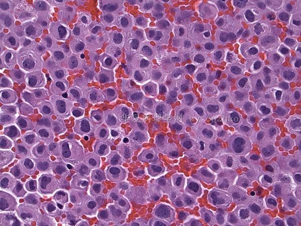
81 y male pleural effusion (cqz-C16)

cqzhao 离线

帖子:5518
粉蓝豆:1071
经验:5755
注册时间:2008-09-29
加关注  |  发消息
楼主 发表于 2010-08-20 22:45|举报|关注(1)
浏览排序[ 顺序 逆序 楼主 支持 精彩 ]  快捷回复
patient has no any malignant history
标签:
0
添加参考诊断
×参考诊断
  

cqzhao 离线

帖子:5518
粉蓝豆:1071
经验:5755
注册时间:2008-09-29
加关注  |  发消息
63 楼    发表于2015-01-14 23:29:47举报|引用
返回顶部 | 快捷回复
引用 62 楼 jani 在 2013-03-24 18:03:18 的发言:

考虑恶性间皮瘤

Thank for your careful review of the case. Can you still have the diagnosis based on the stains

0
回复

cqzhao 离线

帖子:5518
粉蓝豆:1071
经验:5755
注册时间:2008-09-29
加关注  |  发消息
61 楼    发表于2013-03-22 10:36:52举报|引用
返回顶部 | 快捷回复
引用 60 楼 wy1992 在 2013-03-21 20:55:35 的发言:

Thank you Dr Zhao so much that I have learnt a lot from your wonderful case!


You are always welcome

0
回复

cqzhao 离线

帖子:5518
粉蓝豆:1071
经验:5755
注册时间:2008-09-29
加关注  |  发消息
58 楼    发表于2011-08-07 20:17:38举报|引用
返回顶部 | 快捷回复
引用 57 楼 掌心0164 在 2011-08-07 20:07:07 的发言:

欢迎赵老师常来这个版块发帖回帖;不胜感激!!!!

Cngratulation for you as banzhu

0
回复

cqzhao 离线

帖子:5518
粉蓝豆:1071
经验:5755
注册时间:2008-09-29
加关注  |  发消息
56 楼    发表于2011-08-07 19:36:18举报|引用
返回顶部 | 快捷回复
本帖最后由 cqzhao 于 2011-08-07 20:16:19 编辑
引用 55 楼 jani 在 2011-08-02 20:16:56 的发言:
引用 13 楼 viivi薇 在 2010-08-31 09:27:00 的发言:

不排除小圆细胞型恶性间皮瘤的可能性

不除外恶性间皮瘤

Calretinin stain

0
回复

cqzhao 离线

帖子:5518
粉蓝豆:1071
经验:5755
注册时间:2008-09-29
加关注  |  发消息
5 楼    发表于2011-02-05 21:26:00举报|引用
返回顶部 | 快捷回复
以下是引用songmeiying在2010-12-11 16:53:00的发言:

 图1中细胞膜不规则的细胞是凋亡的细胞吗,谢谢。


You mean some mitoses?

0
回复

cqzhao 离线

帖子:5518
粉蓝豆:1071
经验:5755
注册时间:2008-09-29
加关注  |  发消息
6 楼    发表于2010-11-13 22:41:00举报|引用
返回顶部 | 快捷回复
以下是引用与君论道在2010-11-9 19:47:00的发言:

 不在一个级别,不知所云

Sorry
0
回复

cqzhao 离线

帖子:5518
粉蓝豆:1071
经验:5755
注册时间:2008-09-29
加关注  |  发消息
7 楼    发表于2010-09-17 20:14:00举报|引用
返回顶部 | 快捷回复
以下是引用l2530700在2010-9-17 2:32:00的发言:

 看到前面的图片 跟我今天(确切说是昨天)看到一张片子相似,我们这边不能做免疫组化,只能基于形态学报告.要是有空 明天我把图片传传

It is better you do not make definite diagnosis until you are sure. Otherwise you will make mistakes sometimes
0
回复

cqzhao 离线

帖子:5518
粉蓝豆:1071
经验:5755
注册时间:2008-09-29
加关注  |  发消息
8 楼    发表于2010-09-08 08:27:00举报|引用
返回顶部 | 快捷回复

 This case is very special one. Most of the cells are sigle or isolated with same cell population. They have classic features of mesothelial cells. . The key to pick up this case is too many cells. You can imagin It is very easy to miss the case if there are only a few tumor cells present.

Wish every one knows the priciple for pleural or peritoneal fluid cytology: Do some basic stains (Ber-EP4 and calretinin) to distinguish epithelial cells from mesothelial cells if you are not sure the dx.

This is for this case.

Thank people to read and discuss the case, especially Dr. viivi薇 .

0
回复

cqzhao 离线

帖子:5518
粉蓝豆:1071
经验:5755
注册时间:2008-09-29
加关注  |  发消息
9 楼    发表于2010-09-08 08:18:00举报|引用
返回顶部 | 快捷回复

 The tumor cells are positive for ck7 and negative for ck20.

Wish every one knows the diagnosis.

0
回复

cqzhao 离线

帖子:5518
粉蓝豆:1071
经验:5755
注册时间:2008-09-29
加关注  |  发消息
10 楼    发表于2010-09-08 08:16:00举报|引用
返回顶部 | 快捷回复
本帖最后由 于 2010-09-08 08:17:00 编辑  TTF-1

名称:图1
描述:图1
0
回复

cqzhao 离线

帖子:5518
粉蓝豆:1071
经验:5755
注册时间:2008-09-29
加关注  |  发消息
11 楼    发表于2010-09-08 08:14:00举报|引用
返回顶部 | 快捷回复
本帖最后由 于 2010-09-08 08:16:00 编辑  Fprget the cell block photo

名称:图1
描述:图1
0
回复

cqzhao 离线

帖子:5518
粉蓝豆:1071
经验:5755
注册时间:2008-09-29
加关注  |  发消息
12 楼    发表于2010-09-05 20:51:00举报|引用
返回顶部 | 快捷回复
 I will find a time to paste some other stains here
0
回复

cqzhao 离线

帖子:5518
粉蓝豆:1071
经验:5755
注册时间:2008-09-29
加关注  |  发消息
13 楼    发表于2010-09-05 20:50:00举报|引用
返回顶部 | 快捷回复
 Good. TTF-1 is often used for the fluid: it is relative specific marker,  lung and thyroid. Also the lung ca is one of most common ca.
0
回复

cqzhao 离线

帖子:5518
粉蓝豆:1071
经验:5755
注册时间:2008-09-29
加关注  |  发消息
14 楼    发表于2010-09-04 22:02:00举报|引用
返回顶部 | 快捷回复
以下是引用viivi薇在2010-9-4 14:55:00的发言:

 如果只能选择3种标记物,第一个选择TTF-1,它主要表达于甲状腺腺上皮和肺的上皮细胞中

第二个选择是CK7和CK20联合使用标记。

赵老师,谢谢您的家庭作业。这次我的诊断思路正确吗?请赵老师指正。

Very done. I would do these three stains first.

CK7/ck20 stains are very useful for many purposes. We use these two stains for most cases for unknown primary in both cytology and surgical cases.


Should stain TTF1 for this case.

Can you tell us why?

0
回复

cqzhao 离线

帖子:5518
粉蓝豆:1071
经验:5755
注册时间:2008-09-29
加关注  |  发消息
15 楼    发表于2010-09-04 21:59:00举报|引用
返回顶部 | 快捷回复
以下是引用cqzhao在2010-9-3 22:55:00的发言:

以下是引用viivi薇在2010-9-3 13:03:00的发言:

 赵老师,现在是否还可以采用IHC标记物进一步确定原发部位?比如TTF-1,ER等

Why did you want to stain ER for this case?

You did not answer this question.
0
回复

cqzhao 离线

帖子:5518
粉蓝豆:1071
经验:5755
注册时间:2008-09-29
加关注  |  发消息
16 楼    发表于2010-09-04 21:58:00举报|引用
返回顶部 | 快捷回复
以下是引用viivi薇在2010-9-4 14:37:00的发言:

以下是引用cqzhao在2010-9-3 22:54:00的发言:

Seem that only you are interested to this case. We can have one to one talk.

You are right.Suppose that I am your attending and your are my resident (truely you can be my teacher)

Do you think it is a normal case of adenocarcinoma based on the cytology?

thanks,Dr.zhao,I am not only interested that case,I want to learn knowledge of cytopathology from this case.

I am not sure if it is a normal case of adenocarcinoma,maybe we can make some ICC makers for it to find a correct result.

It is unusual case. The cells look like mesothelial cells, but they are not
0
回复

cqzhao 离线

帖子:5518
粉蓝豆:1071
经验:5755
注册时间:2008-09-29
加关注  |  发消息
17 楼    发表于2010-09-03 23:04:00举报|引用
返回顶部 | 快捷回复

 viivi薇 : After we know this is a metastatic ca,  we must continue to work on the case and try to figure out the origin of the tumor. It is our duty to provide the correct and detailed information to clinicians and patients. Of cause often we cannot tell the exact origin in clinical practice. However  we should try to or at least give some clues or suggestion.

Question No. 2

If you can only do three stains, tell us what three markers you will stain? I will come here to check the home work.   If others want to make your suggestion you are welcome too.

 thanks, cz

0
回复

cqzhao 离线

帖子:5518
粉蓝豆:1071
经验:5755
注册时间:2008-09-29
加关注  |  发消息
18 楼    发表于2010-09-03 22:55:00举报|引用
返回顶部 | 快捷回复
以下是引用viivi薇在2010-9-3 13:03:00的发言:

 赵老师,现在是否还可以采用IHC标记物进一步确定原发部位?比如TTF-1,ER等

Why did you want to stain ER for this case?
0
回复

cqzhao 离线

帖子:5518
粉蓝豆:1071
经验:5755
注册时间:2008-09-29
加关注  |  发消息
19 楼    发表于2010-09-03 22:54:00举报|引用
返回顶部 | 快捷回复
以下是引用viivi薇在2010-9-3 12:35:00的发言:

 谢谢赵老师的指正,关于Calretinin and Ber-EP4在ICC标记物中的意义,我是查找国内的一本参考资料--《免疫组织化学病理诊断》P90中的表8-2找到的,谢谢赵老师告诉我这两种标记物正确的意义所在,现在从免疫组化的结果考虑转移性腺癌,赵老师,这次我对了吗?

Seem that only you are interested to this case. We can have one to one talk.

You are right.Suppose that I am your attending and your are my resident (truely you can be my teacher)

Do you think it is a normal case of adenocarcinoma based on the cytology?

0
回复

cqzhao 离线

帖子:5518
粉蓝豆:1071
经验:5755
注册时间:2008-09-29
加关注  |  发消息
20 楼    发表于2010-09-02 20:23:00举报|引用
返回顶部 | 快捷回复

 Thank your explanation.


如果整张涂片观察Calretinin呈局灶阳性,Ber-EP4强阳性,就考虑肺腺癌;如果整张涂片Calretinin为阴性,则考虑乳腺癌. Above is not correct.

Both lung and breast cancers (almost all epithelial tumors) are positive for Ber-EP4 and negative for calretinin. Ber-EP4 and calretinin are only used for distinguish epithelial cells from mesothelial cells. They cannot tell the origins and the nature of the cells (benign or malignant). Of cause generally speaking if you find epithelial cells in 腹水 或pleural fluid are metastatic tumors.

Above are basic stain priniciple in fluid IHC. for your reference

0
回复
回复:63 阅读:10517
共2页/29条首页上一页12下一页尾页
【免责声明】讨论内容仅作学术交流之用,不作为诊疗依据,由此而引起的法律问题作者及本站不承担任何责任。
快速回复
进入高级回复
您最多可输入10000个汉字,按 "Ctrl" + "Enter" 直接发送
搜索回复/乘电梯 ×
按内容
按会员
乘电梯
合作伙伴
友情链接