| 图片: | |
|---|---|
| 名称: | |
| 描述: | |
- 子宫内膜
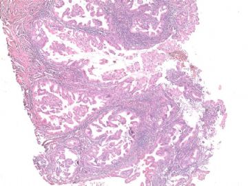
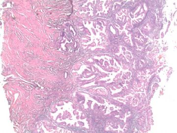
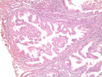
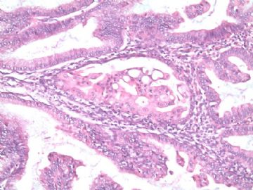
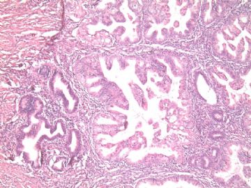
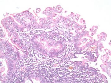
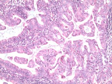
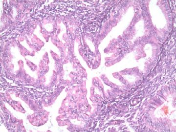
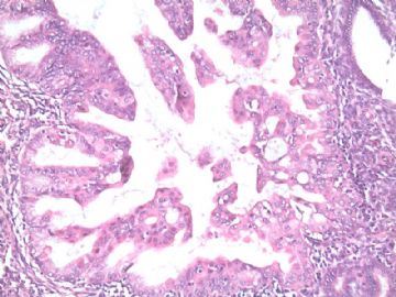
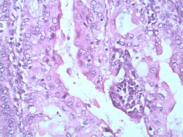
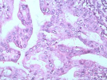
The H&E sections showc omplex papillary structure and the cells wtih mild to moderate cytologic atypia. At least it is atypical complex hyperplasia.
是不是内膜内癌就见人见智. It is true for this case. It is better to read the glass slides and know the size of focal atypical proliferation.
Based on the cytomorphology and negative P53 stain, it is not a type 2 tumor.
I favor a dx of focal endometrial adenocarcinoma, endometrioid type, FIGO grade 1, in the background of atypical complex hyperplasia.
Just for your reference.
沿金老师的思路考虑鉴别诊断:
1 良性和化生
乳头状全体细胞化生和嗜酸性化生见于表面上皮。鳞化可见于表面上皮,也见于腺体(桑葚)。它们均无真正的核非典型性。A-S反应有相应的间质改变并且与周围腺体存在移行过渡。本例不属于上述任何一种情形。
2 不典型复杂性增生
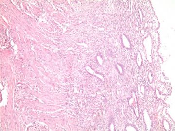
真正的核非典型性判断(请仔细观察图6,对比病变与周围正常腺体体会结构异常,观察最后几图体会核异常):核增大、变圆、染色质空亮、出现明显核仁。核级别已达到中-高级。此时如果是内膜癌,分级上升1级。
3 内膜样癌(IA期)
IA期指癌局限于内膜内,即:内膜内癌,或子宫内膜间质浸润,但无肌层浸润。
判断子宫内膜间质内浸润具有一定主观性,在此不作勉强区分。再观察图6,病变与周围正常腺体相穿插,是否认为是间质内浸润的一种表现呢?
总之,本例有肯定的细胞学异型性和结构异常,至少为复杂性不典型增生。如果是我的病例,报子宫内膜癌(2级,IA期)。

华夏病理/粉蓝医疗
为基层医院病理科提供全面解决方案,
努力让人人享有便捷准确可靠的病理诊断服务。
