
图1")
图2")
图3")
图4")
图5")
图6")
图7")
图8")
图9")
图10")
图11")
图12")
图13")
图14")

 好病例,赞同Dr.cqzhao,考虑LCIS并累及硬化性腺病。


| 图片: | |
|---|---|
| 名称: | |
| 描述: | |
- B2770女/38岁, 右侧乳腺穿刺 诊断?23楼-手术标本(2010-7-24)
图1") 图1
图1图2") 图2
图2图3") 图3
图3图4") 图4
图4图5") 图5
图5图6") 图6
图6图7") 图7
图7图8") 图8
图8图9") 图9
图9图10") 图10
图10图11") 图11
图11图12") 图12
图12图13") 图13
图13图14") 图14
图14
| 姓 名: | ××× | 性别: | 年龄: | ||
| 标本名称: | |||||
| 简要病史: | 右侧乳腺钼靶示2cm 左右不规则肿块,无钙化。PET/CT示高度代谢。 | ||||
| 肉眼检查: | 穿刺组织8条。 | ||||
标签:LCIS 硬化性腺病
-
本帖最后由 于 2010-07-24 13:18:00 编辑

- xljin8
相关帖子
- • 左乳腺肿物,请教诊断?
- • 乳腺肿物
- • 左乳肿物
- • 乳腺肿物一例
- • 腺病?癌?其他?(12楼常规,24楼免疫组化及会诊结果)
- • 乳腺肿物
- • 乳腺癌?
- • 求助:56岁女,左乳肿物,能排除小管癌吗?
- • 38岁乳腺(新加HE切片)
- • 乳腺肿块
×参考诊断
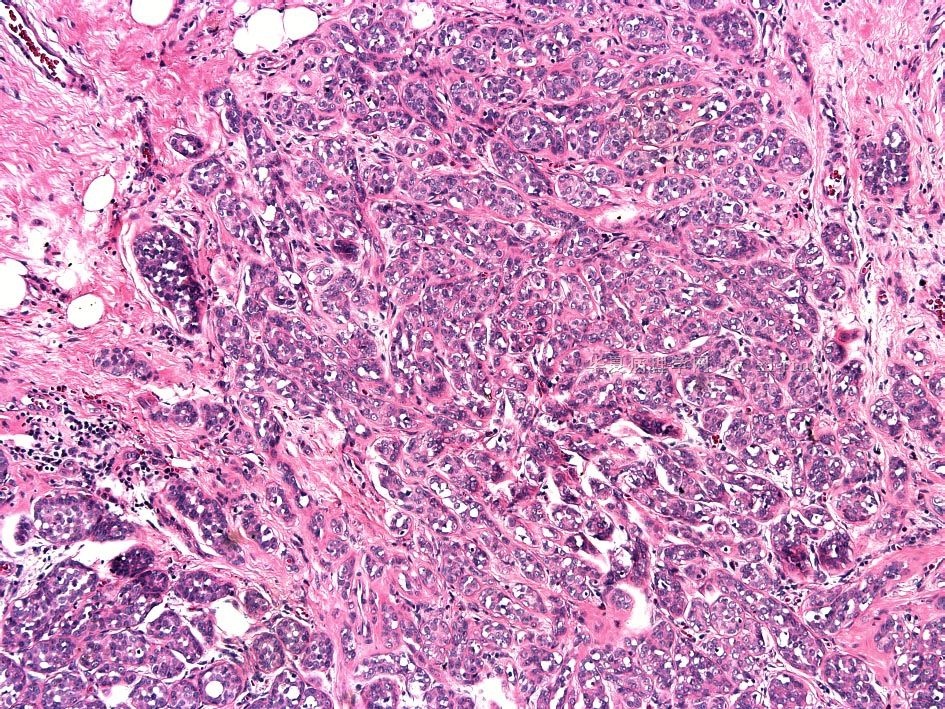
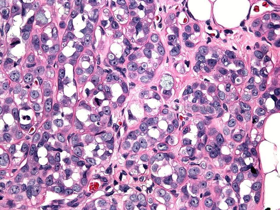
活检:硬化性乳腺病伴小叶不典型增生,未能除外小叶原位癌。手术标本:小叶原位癌累及硬化性腺病?
以点看线,以线看面,以面推全的不成熟的看法:
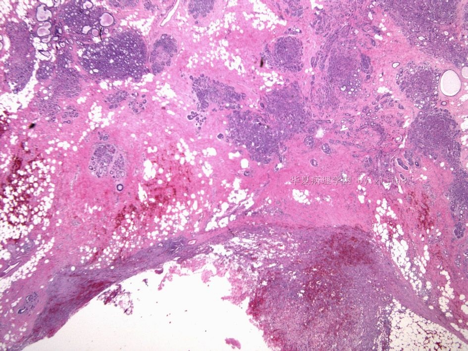
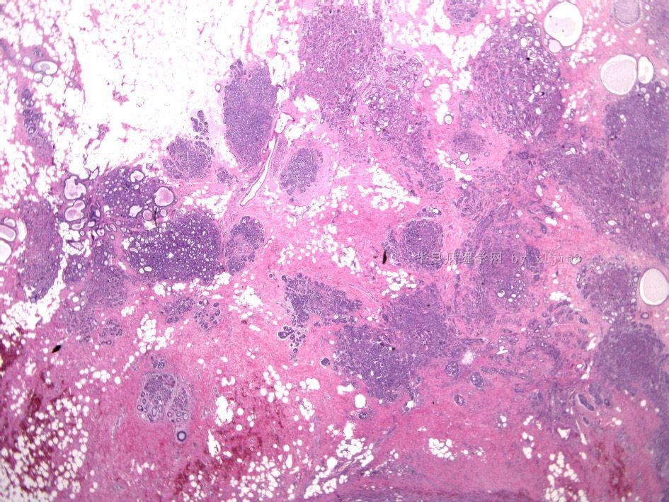
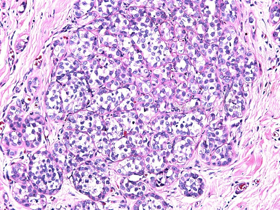
新增的乳腺切除标本,低倍显示边缘脂肪组织及乳腺小叶的出血区,提示可能为穿刺部位周边,但未见明显炎症反应。
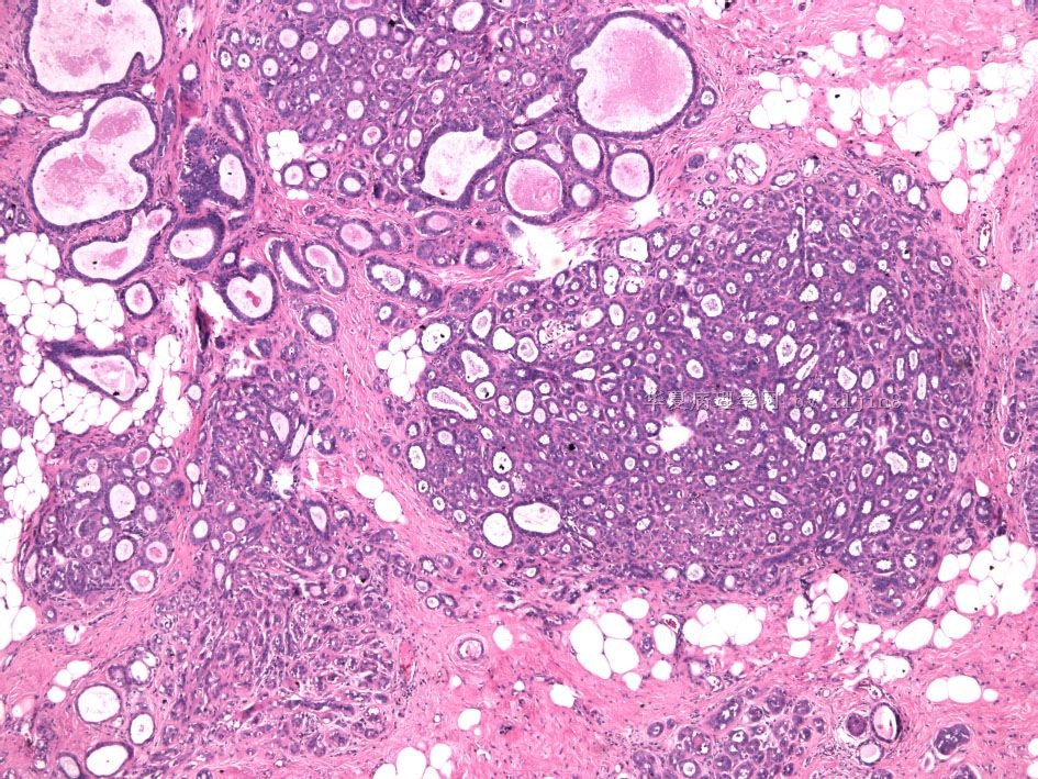
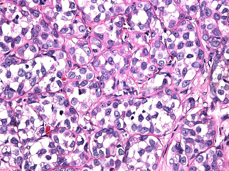
大部分的病变特点与穿刺活检标本边缘区的病变相似为小叶增生性病变。但本次标本的图5,显示该小叶大致轮廓尚存,但小叶内腺管增生明显并具一定非典型性,表现为较小的管样、条索样、不规则样其间可见硬化的间质(可能为伴有小叶非典型增生的硬化性腺病组织像,是否可考虑为小叶原位癌可能?)。
本例,Dr. XLJin8必定大量取材,多切面观察,但是仍未见与穿刺活检 焦点图片完全类似的区域,期待能有新的发现。
谢谢您的病例,收藏期待后期病人随访情况的报道。
-
本帖最后由 于 2010-07-28 19:41:00 编辑
| 以下是引用cqzhao在2010-7-24 21:39:00的发言:
Often it is difficult to distinguish ALH from LCIS even though there are some definitions. In fact both ALH and LCIS represent the similar meaning----the risk fact of invasive carcinoma in both sides of breast. We will nnot report the margins for both. For this case, extensive lobular lesion involves sclerosing adenosis. So it is good to diagnose as LCIS with involvement of sclerosing adnosis. The patient need to have clinical following up with imaging. |
Often it is difficult to distinguish ALH from LCIS even though there are some definitions. In fact both ALH and LCIS represent the similar meaning----the risk fact of invasive carcinoma in both sides of breast. We will nnot report the margins for both.
For this case, extensive lobular lesion involves sclerosing adenosis. So it is good to diagnose as LCIS with involvement of sclerosing adnosis. The patient need to have clinical following up with imaging.
-
CHENYINQIAO 离线

- 帖子:560
- 粉蓝豆:0
- 经验:606
- 注册时间:2010-07-20
- 加关注 | 发消息
请关注以下病例,与本例好像有相似又有不同。
http://www.ipathology.cn/forum/forum_display.asp?keyno=276829
尝试翻译,请参考
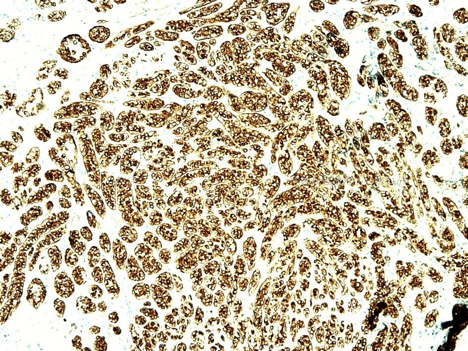
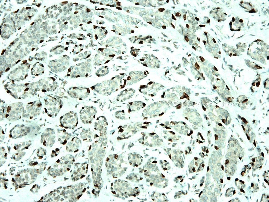
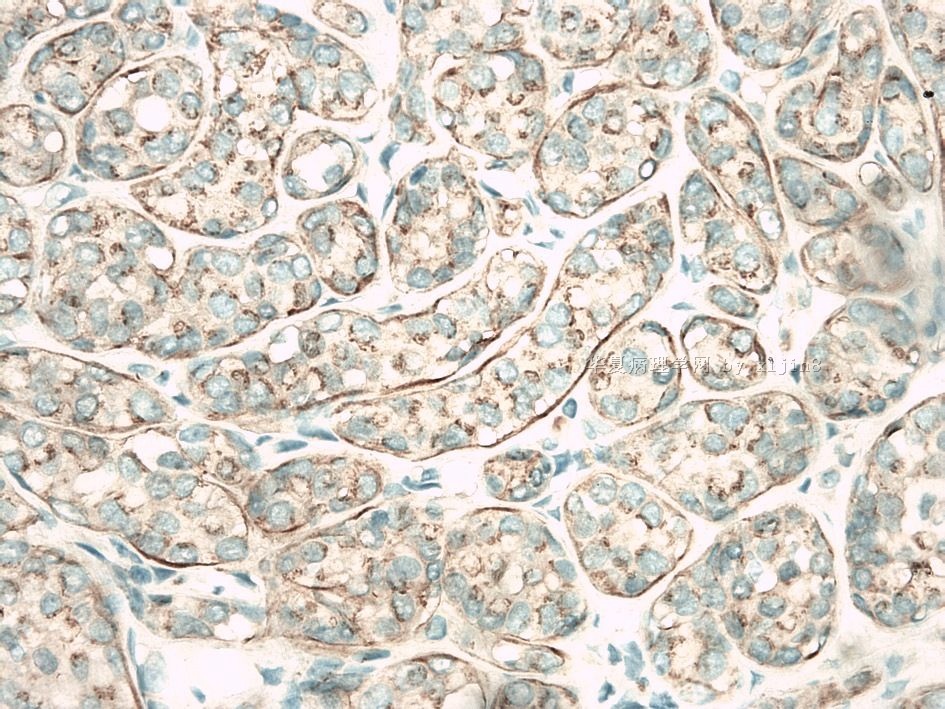
E-cad expression is reduced based on the stains above.
基于以上的染色结果显示E-cadherin表达减少。
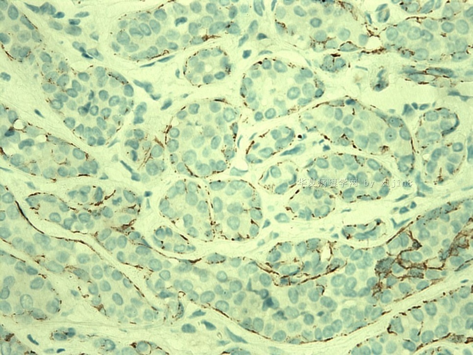
The key for this case to rule out invasice component.
对于本例重点是找出浸润性的成分。
If area for p63 stain is above area. No invasion is present
如果P63染色是以上的区域。那么没有浸润的证据。
Atypical lobular hyperplasia involving sclerosing adenosis.
包含于硬化性腺病中的非典型性小叶增生。


