


| 图片: | |
|---|---|
| 名称: | |
| 描述: | |
- 胸水液基细胞学-地坛
Feeling these cell clusters are the same cell population and the cytologic features are not very urgly. It is bettter to know the previous breast type and see the slides.
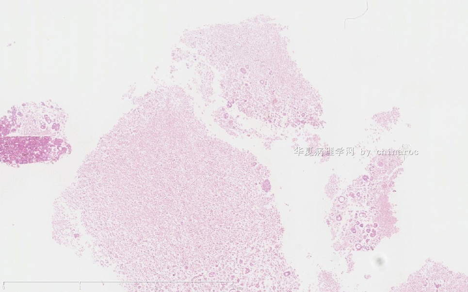
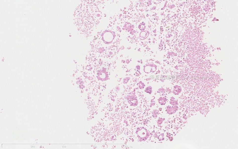
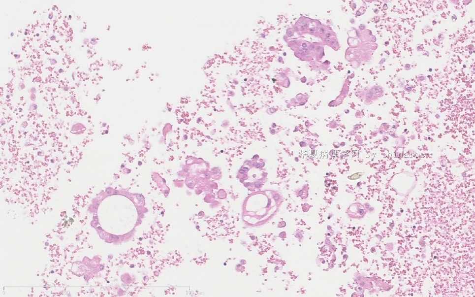
These cells show round uniform nuclei with abundent cytoplasma. Question is that they are mesothelial cells or epithelial cells. If they are epithelial cells, they are metastatic tumor cells.
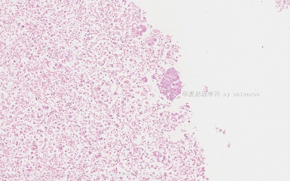
For this case the cellularity is very high, so it is easy to have a cell block. You can stain berep4 and calretinin. It is easy to get answer.
For fluid cytology we always make cell block. For unsure cases, we always do epithelial and mesothelial marker stains.
-
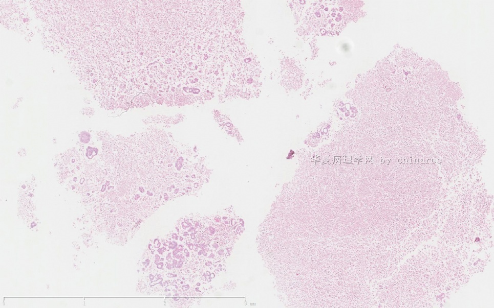
Thanks for Dr. Zhao's analysis about this case. To me it is difficult to make a definite diagnosis at first eye. At some points I finally give a positive diagnosis of malignancy. The cellularity which Dr. Zhao has referred is the most important criteria. The another one is the architecture forming glandular particles with little lymphocytes infiltration.
According to Zhao's advice, several markers such like CK5/6, calretinin and mesothelia will be done based on cell block.

- 用心做事、真情做人!
-
Feeling these cell clusters are the same cell population and the cytologic features are not very urgly. It is bettter to know the previous breast type and see the slides.
These cells show round uniform nuclei with abundent cytoplasma. Question is that they are mesothelial cells or epithelial cells. If they are epithelial cells, they are metastatic tumor cells.
For this case the cellularity is very high, so it is easy to have a cell block. You can stain berep4 and calretinin. It is easy to get answer.
For fluid cytology we always make cell block. For unsure cases, we always do epithelial and mesothelial marker stains.
感觉这些细胞簇是相同的细胞群体,且其细胞学特征不清晰。最好了解以前的乳腺癌类型并阅片。
这些细胞为圆形均一核伴大量胞浆。问题是他们是间皮细胞还是上皮细胞?如果是上皮细胞,则是转移性肿瘤细胞。
对于该例,细胞很丰富,因此应该比较容易得到细胞块。可以染BerEP4和calretinin,很容易得到答案。
对于液基细胞,我们都做细胞块。对于不确定病例,一般做上皮和间皮标记的染色
- 赚点散碎银子养家,乐呵呵的穿衣吃饭
| Thanks for Dr. Zhao's analysis about this case. To me it is difficult to make a definite diagnosis at first eye. At some points I finally give a positive diagnosis of malignancy. The cellularity which Dr. Zhao has referred is the most important criteria. The another one is the architecture forming glandular particles with little lymphocytes infiltration. According to Zhao's advice, several markers such like CK5/6, calretinin and mesothelia will be done based on cell block. | ||
|
谢谢Dr.Zhao对该例的分析。对我来讲,第一眼很难做出确定的诊断。根据某些方面,我最终给出了恶性的阳性诊断。Dr.Zhao指出的其细胞构成是最重要的参考。另一方面是其形成腺样结构,伴少量淋巴细胞浸润。 根据Dr.Zhao的建议,将在细胞块上座一些标记
|
- 赚点散碎银子养家,乐呵呵的穿衣吃饭
-
Best epithelial marker is BerEp4. Based on patient's history you also can stain ER.
Pathologists need to answer the questions for plural fluid exam:
Reactive or metastatic?
Origins or primary site if it is metastatic tumor?
For women most common primary sites include lung, breast, gynecologic orgains.
最好的上皮标记是BerEp4。根据患者病史,还应染ER。
病理工作者对于胸膜液基标本需要回答以下问题:
反应性还是转移性?如果是转移性肿瘤那么其来源或原发位置?对于女性,最常见的原发位置有肺、乳腺、生殖道
- 赚点散碎银子养家,乐呵呵的穿衣吃饭
The bad sign of cytology for this case is very celluar specimen with the same cell population and bloody fluid.
Interesting to know if the patient had lobular ca.
Anyway i will do some stains before I sign out the case.
对于该例,恶性的细胞学特征为相同的细胞群组成的丰富的细胞标本、血性。
很想知道该患者是否为小叶癌
总之在签发该例之前我会做标记
- 赚点散碎银子养家,乐呵呵的穿衣吃饭
Best epithelial marker is BerEp4. Based on patient's history you also can stain ER.
Pathologists need to answer the questions for plural fluid exam:
Reactive or metastatic?
Origins or primary site if it is metastatic tumor?
For women most common primary sites include lung, breast, gynecologic orgains.
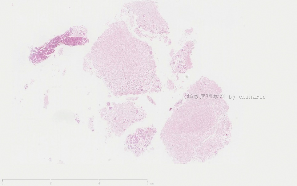
| 以下是引用cqzhao在2010-7-16 3:46:00的发言: Cell block shows glandular cells. So dx of metastatic ca should not have problem. But pathologists need to stain and provide more information for the primary. |
cqzhao老师回复
细胞块显示腺细胞。因此转移癌的诊断应当无问题。但我们需要做染色并提供其他原发疾病的信息
- 赚点散碎银子养家,乐呵呵的穿衣吃饭
-
CHENYINQIAO 离线

- 帖子:547
- 粉蓝豆:0
- 经验:593
- 注册时间:2010-07-20
- 加关注 | 发消息


