| 图片: | |
|---|---|
| 名称: | |
| 描述: | |
- 颈部肿物
-
本帖最后由 于 2008-02-19 12:23:00 编辑
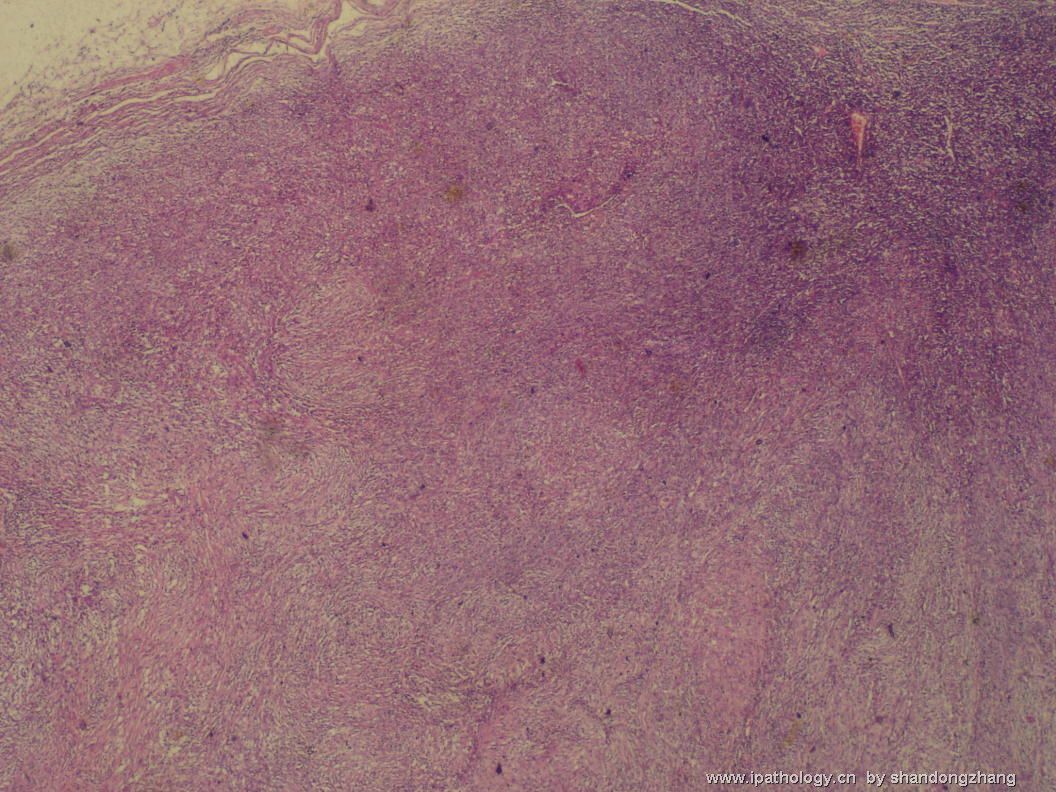
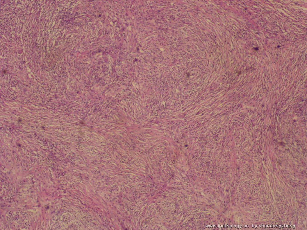
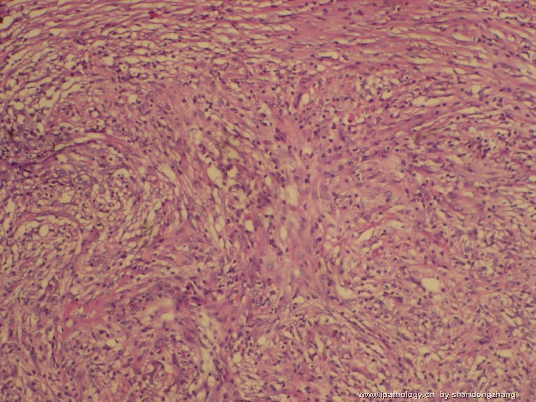
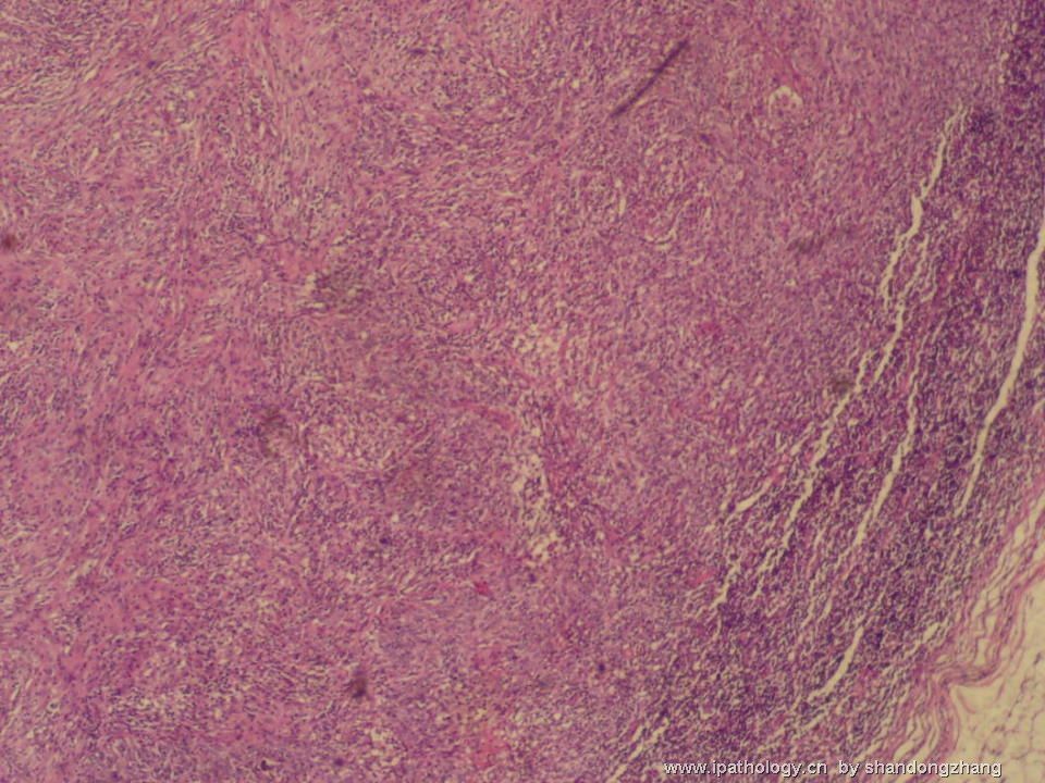
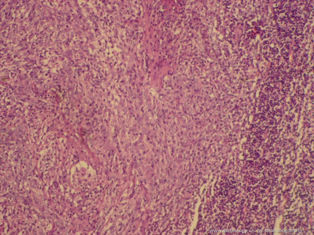
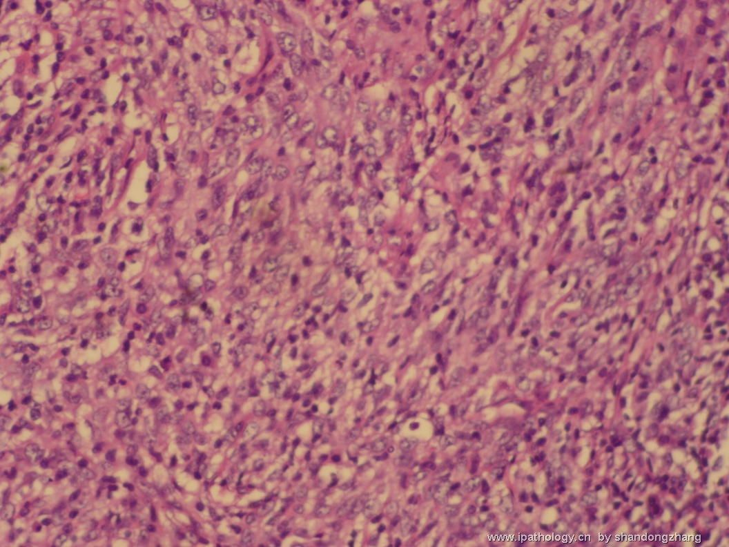
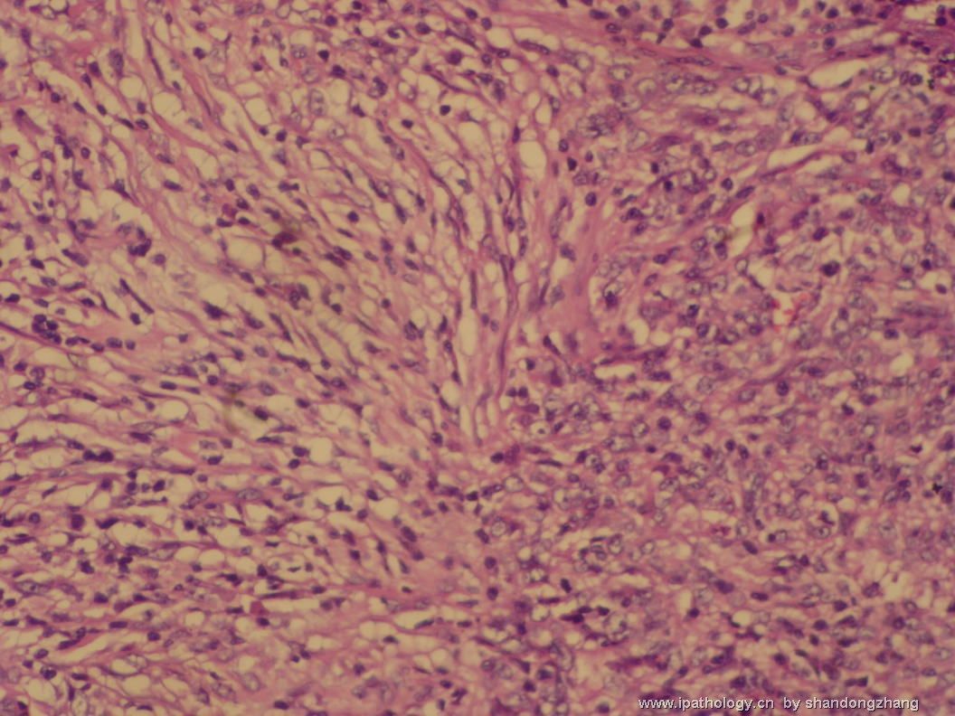
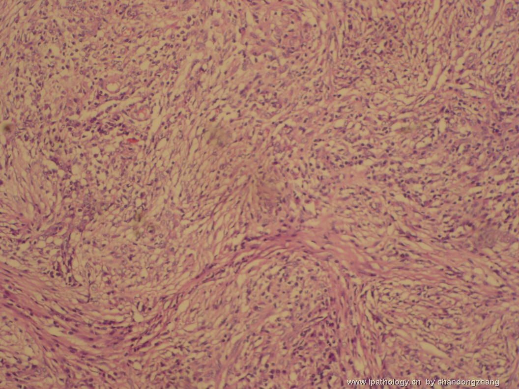
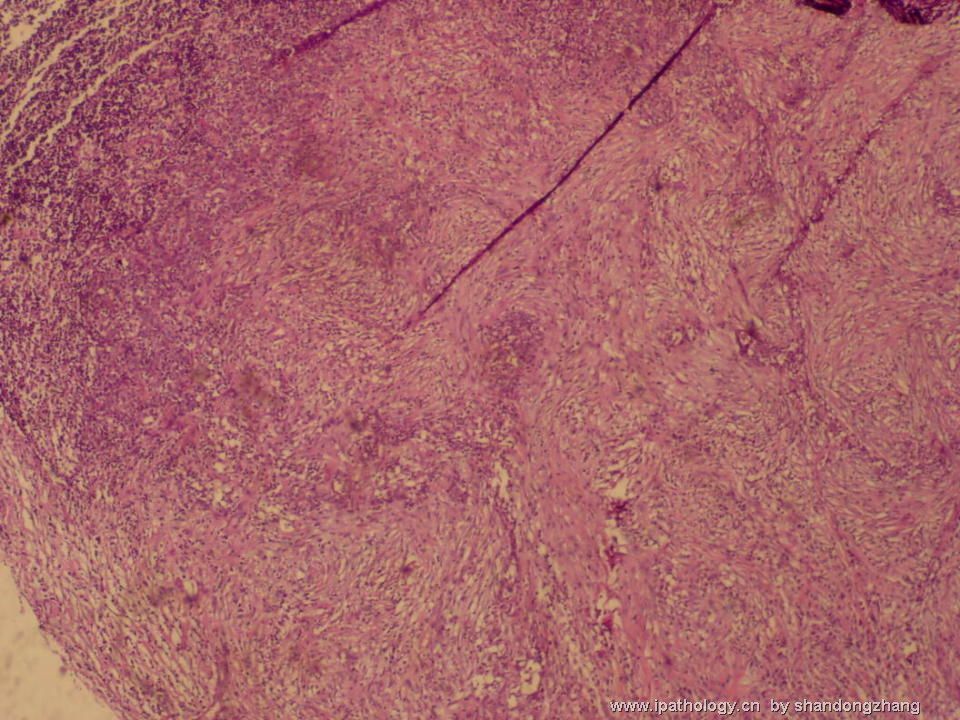
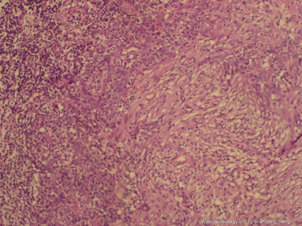
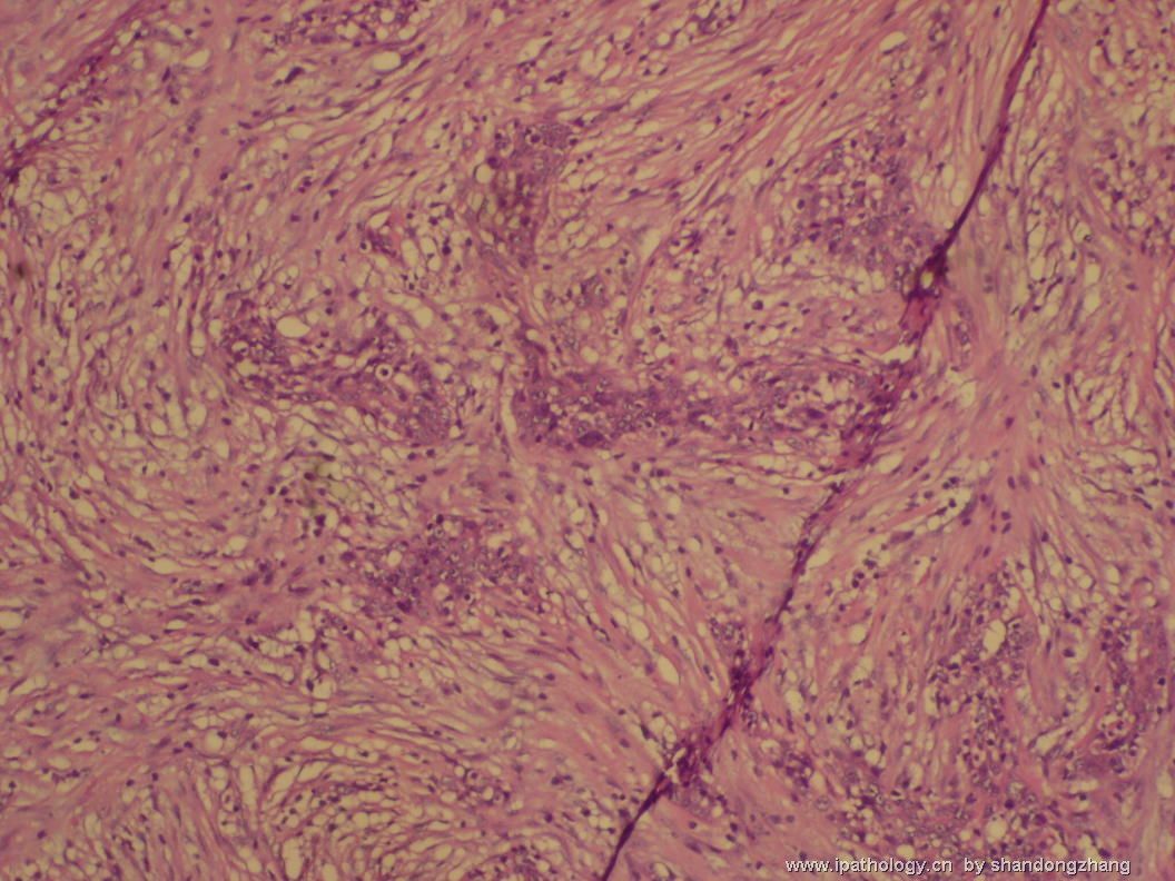
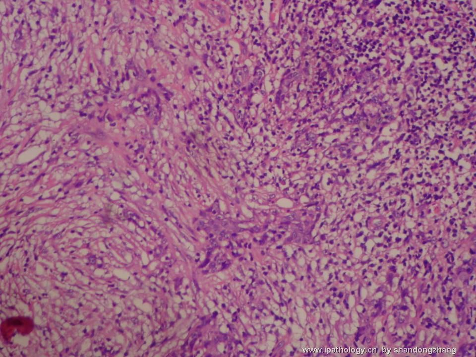
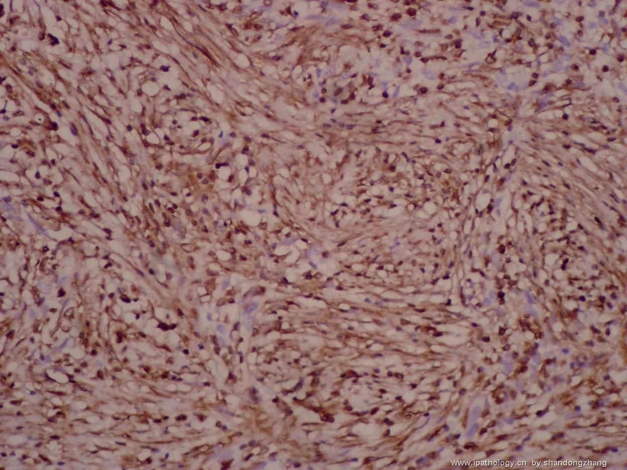
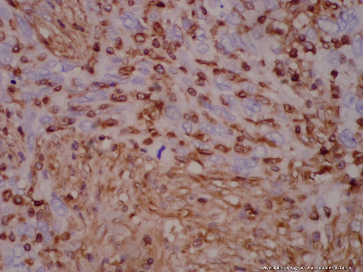
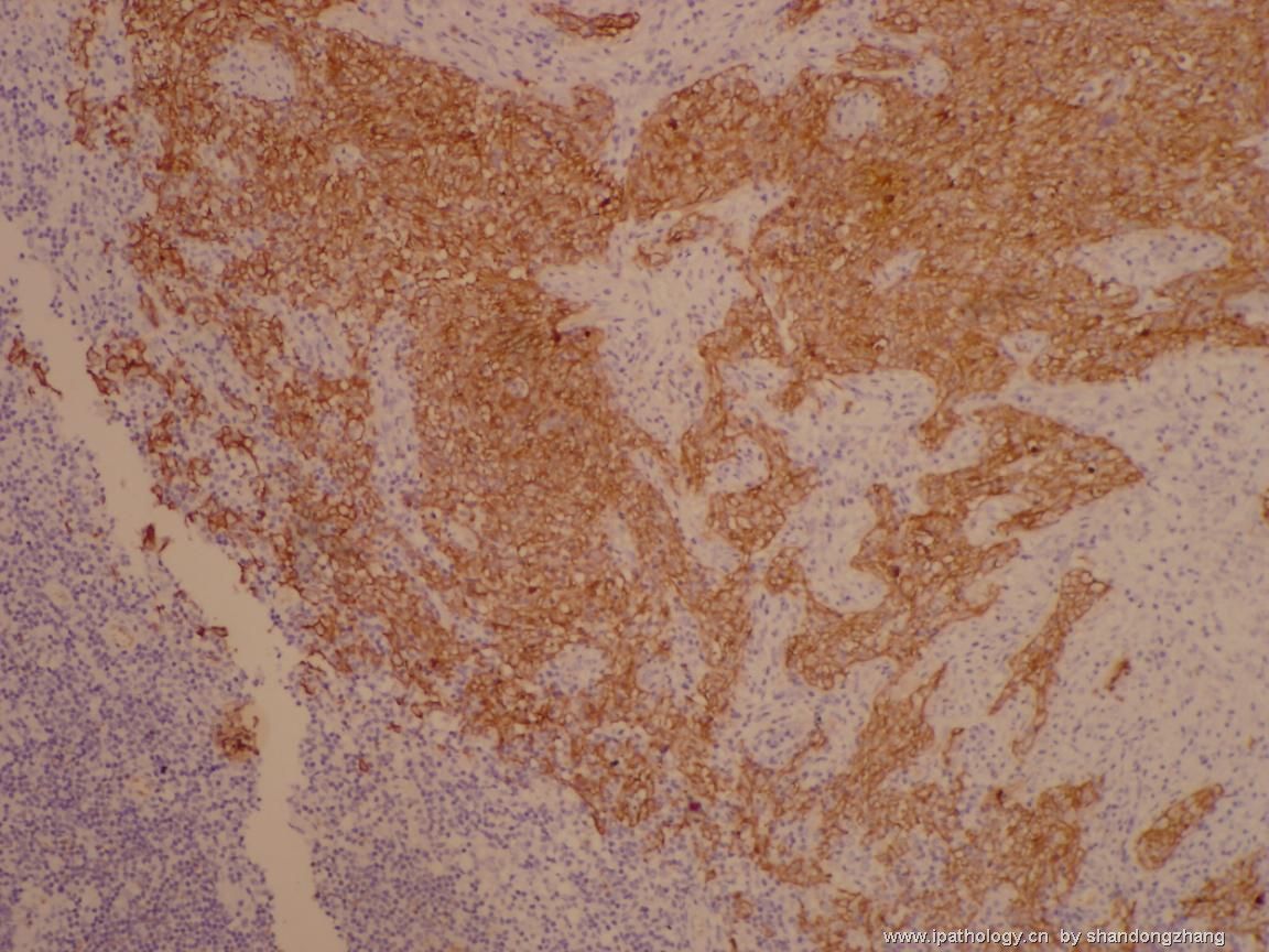
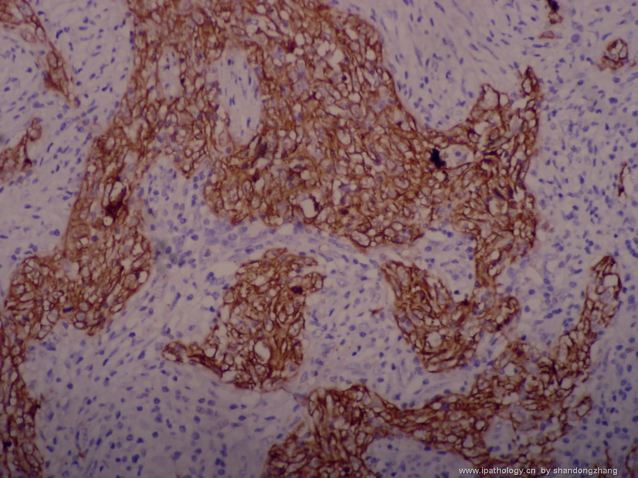
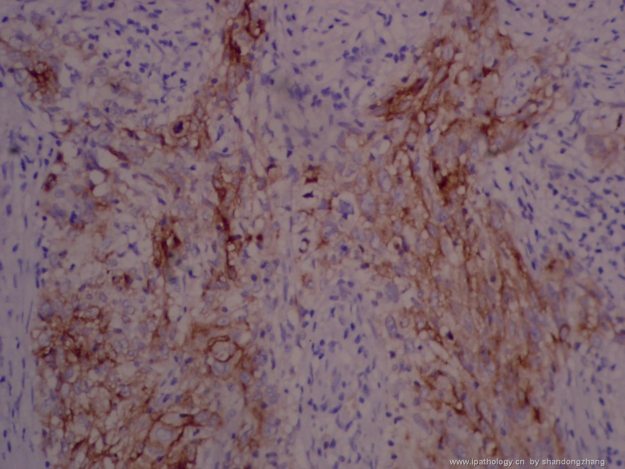
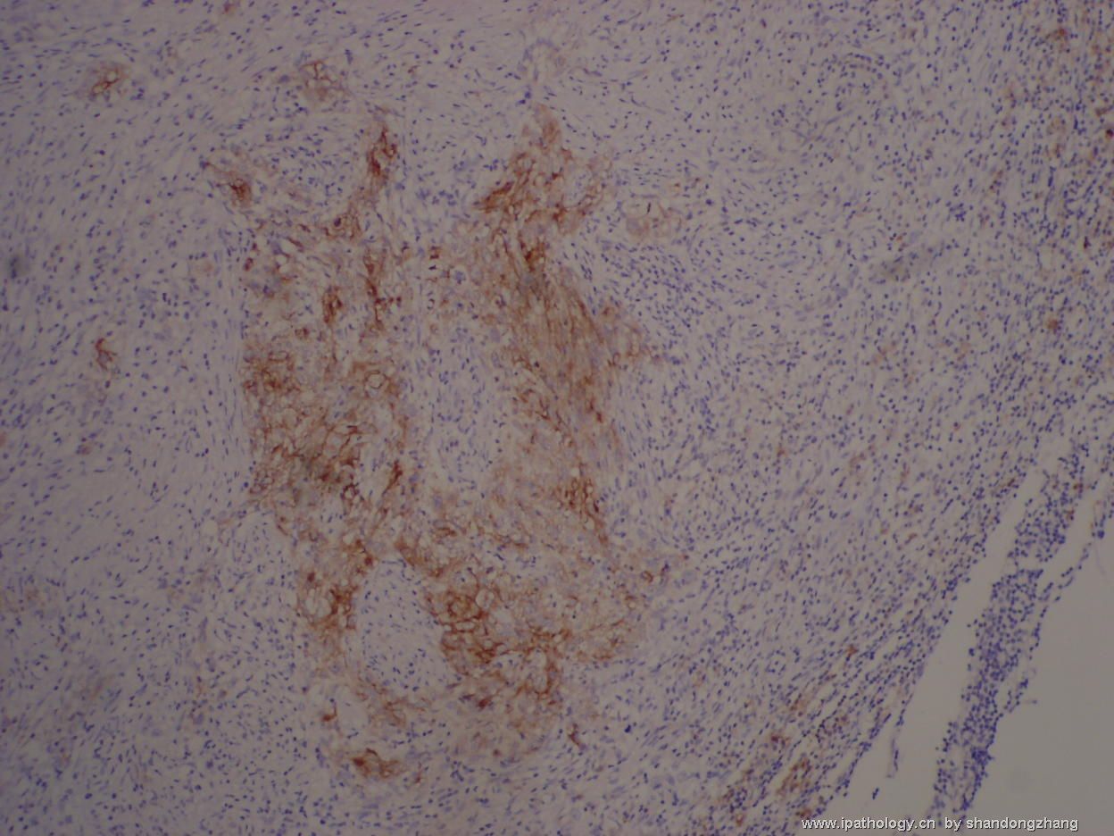
淋巴结树突状细胞肿瘤并非一定CK或者EMA阴性,老年人颈部淋巴结肿大当然首先除外转移癌,但FDC, IDC,CIRC也可以表达CK或者EMA,此例但从形态上看不除外这些肿瘤,前提是进一步检查鼻咽部,重新做CD21和CD35等。以下是几篇参考文献摘要:
AIMS: Tumours of dendritic/accessory cell origin are rare neoplasms arising in lymph nodes. Among these, tumours derived from cytokeratin-positive interstitial reticulum cells (CIRCs), a subset of fibroblastic reticulum cells, are reported even less frequently. The International Lymphoma Study Group (ILSG) has recently proposed a classification for tumours of histiocytes and accessory dendritic cells in which CIRC tumours are not included. We report a case of a CIRC tumour arising in a submandibular lymph node of a 66-year-old male. METHODS AND RESULTS: The neoplasm was composed of spindle cells with elongated or round nuclei, prominent nucleoli and abundant cytoplasm. These cells were arranged in a diffuse fascicular and vaguely whorled pattern. The tumour cells stained diffusely for S100, vimentin, desmin, lysozyme, and focally for CD68 and cytokeratins 7, 8, 18, CK-AE1 and CK-pool. Electron microscopy was performed for further evaluation on samples taken from the paraffin block; this revealed cytoplasmic projections and rudimentary cell junctions. CONCLUSIONS: Histopathologist should be aware of the existence of tumours deriving from CIRCs, as these cases may be misdiagnosed as metastatic carcinoma. Careful clinical and pathological evaluation is necessary to exclude this possibility.
BACKGROUND: Follicular dendritic cell (FDC) tumors are rare. A majority of the reported cases were confined to the lymph nodes. We report a case of FDC tumor occurring in the parapharyngeal region in a 45-year-old woman. METHODS: Characteristic histopathologic features of the excised primary and recurrent parapharyngeal tumors in conjunction with immunohistochemistry and electron microscopy helped us to arrive at a diagnosis of FDC tumor. RESULTS: Histopathology of primary excision revealed a lobulated tumor with a suggestion of ill-defined whorls. The most striking feature was regular occurrence of aggregates of lymphocytes within the tumor, especially around the blood vessels. The anatomic location together with the histology indicated the possibilities of either a meningioma, a salivary gland tumor, or a nerve sheath tumor. Immunostains for cytokeratin (CK), S-100 protein, and smooth muscle actin (SMA) were negative. However, the tumor cells showed strong immunoreactivity for epithelial membrane antigen (EMA) and vimentin. A diagnosis of parapharyngeal meningioma appeared to be the closest possibility. One year later, the patient developed a recurrence at the same site. A reexcision showed an identical tumor with an additional feature of lymphatic embolization and angioinvasion. A review of the entire case with further immunoreactivity for CD21 and CD35 confirmed the diagnosis of FDC. CONCLUSIONS: Follicular dendritic cell tumor has distinctive morphologic features and immunohistochemical profile. It is also characterized by considerable potential for recurrences.

- the more we discuss, the more we learn from each other !!
| 以下是引用Chiang在2007-9-12 5:50:00的发言: 头颈部梭形细胞肿瘤尤其是老年人首先考虑梭形细胞癌(肉瘤样癌),诊断需要免疫标记证实其上皮表达,同时除外血管源性,需要注意的是梭形细胞癌vimentin常阳性表达,而且CK表达和很少,甚至有的种类不表达,常用的标记如Cam 5.2,AE1/3等,有些肉瘤样癌可呈假血管肉瘤样,因此,应标记血管内皮,阴性者支持癌,而单独一个CK表达不能肯定为癌,血管肉瘤可以表达CK。 |

- the more we discuss, the more we learn from each other !!
-
caotong_1978 离线
- 帖子:286
- 粉蓝豆:71
- 经验:698
- 注册时间:2006-12-13
- 加关注 | 发消息
-
lfl001200546 离线
- 帖子:2808
- 粉蓝豆:40
- 经验:2808
- 注册时间:2007-02-14
- 加关注 | 发消息
-
liguoxia71 离线
- 帖子:4174
- 粉蓝豆:3122
- 经验:4677
- 注册时间:2007-04-01
- 加关注 | 发消息
IDCS?ck+?不太可能啊
IDCS:Immunophenotype
o S-100 +
o Negative for vimentin and CD1a
o Variably, weakly + for CD68, lysozyme, and CD45
o Negative for FDCs (CD21 and CD35), MPO, CD34, specific B and T-cell antigens, CD30, CK, EMA
猜一个:转移性肉瘤样癌

知之者不如好之者,好之者不如乐之者。(语出幽梦影)

