共1页/15条首页上一页1下一页尾页
回复:15 阅读:4185
舌缘肿块,上皮内瘤变分级?

太行山 离线

帖子:115
粉蓝豆:425
经验:304
注册时间:2010-06-09
加关注  |  发消息
楼主 发表于 2010-06-11 12:46|举报|关注(0)
浏览排序[ 顺序 逆序 楼主 支持 精彩 ]  快捷回复
姓    名: ××× 性别:  男 年龄:  62
标本名称:  舌缘肿块
简要病史:  舌缘白色斑块两月余,无明显不适。
肉眼检查:  灰白色组织1X0.7XO.5
  • 舌缘肿块,上皮内瘤变分级?图1
    图1
  • 舌缘肿块,上皮内瘤变分级?图2
    图2
  • 舌缘肿块,上皮内瘤变分级?图3
    图3
  • 舌缘肿块,上皮内瘤变分级?图4
    图4
  • 舌缘肿块,上皮内瘤变分级?图5
    图5
  • 舌缘肿块,上皮内瘤变分级?图6
    图6
标签:
本帖最后由 于 2010-06-12 09:33:00 编辑
0
×参考诊断
鳞状上皮轻-中度非典型增生,建议临床随访.

wfbjwt 离线

帖子:6628
粉蓝豆:163
经验:7418
注册时间:2006-10-08
加关注  |  发消息
1 楼    发表于2010-06-11 18:45:00举报|引用
返回顶部 | 快捷回复
 呵呵,CIN只用于宫颈。此例基本够鲍温病了。
0
回复
signature
嫁人就嫁灰太狼,学习要上华夏网。

太行山 离线

帖子:115
粉蓝豆:425
经验:304
注册时间:2010-06-09
加关注  |  发消息
2 楼    发表于2010-06-12 10:52:00举报|引用
返回顶部 | 快捷回复
 谢谢wfbjwt老师,已更正。
0
回复

海上明月 离线

帖子:9476
粉蓝豆:1172
经验:10007
注册时间:2009-08-29
加关注  |  发消息
3 楼    发表于2010-06-13 07:28:00举报|引用
返回顶部 | 快捷回复
 鳞状上皮中-重度异型增生,局灶重度异型增生(符合原位鳞癌)。
0
回复
signature
王军臣

xclbljys 离线

帖子:5253
粉蓝豆:224
经验:7005
注册时间:2008-11-21
加关注  |  发消息
4 楼    发表于2010-06-13 09:42:00举报|引用
返回顶部 | 快捷回复
本帖最后由 于 2010-06-13 09:58:00 编辑
以下是引用海上明月在2010-6-13 7:28:00的发言:

 鳞状上皮中-重度异型增生,局灶重度异型增生(符合原位鳞癌)。


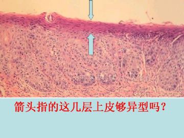
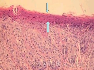
海上明月老师:发生在舌部的上皮内瘤变,其组织学特点是否与宫颈有所不同?我观察本例最重的病变部分(图4),表面似乎仍有数层成熟细胞的存在,这种情况我甚至不敢报重度异型增生,请老师给予指导!

0
回复
signature
许春雷

xclbljys 离线

帖子:5253
粉蓝豆:224
经验:7005
注册时间:2008-11-21
加关注  |  发消息
5 楼    发表于2010-06-13 09:53:00举报|引用
返回顶部 | 快捷回复
请老师观察箭头所指!
  • 图1
0
回复
signature
许春雷

xclbljys 离线

帖子:5253
粉蓝豆:224
经验:7005
注册时间:2008-11-21
加关注  |  发消息
6 楼    发表于2010-06-13 10:04:00举报|引用
返回顶部 | 快捷回复
我把上图再放大一点。
  • 图1
0
回复
signature
许春雷

水中鱼儿 离线

帖子:550
粉蓝豆:13
经验:924
注册时间:2010-03-26
加关注  |  发消息
7 楼    发表于2010-06-13 23:08:00举报|引用
返回顶部 | 快捷回复
 口腔粘膜上皮内瘤变(OIN)不知是否包含舌,如果包含舌的话,即使是低级别也是很危险的,相当于其他部位的高级别危险性。
0
回复

广秀 离线

帖子:2711
粉蓝豆:1246
经验:3502
注册时间:2009-09-15
加关注  |  发消息
8 楼    发表于2010-06-20 06:23:00举报|引用
返回顶部 | 快捷回复
 感觉箭头所指是表面的角化不全及角化过度层,报中-重度不典型增生,部分区域有原位癌的趋势。
0
回复
signature
人淡如菊,心素如简。

XLJin8 离线

帖子:2275
粉蓝豆:1531
经验:2417
注册时间:2009-03-04
加关注  |  发消息
9 楼    发表于2010-06-20 22:14:00举报|引用
返回顶部 | 快捷回复
本帖最后由 于 2010-06-21 10:32:00 编辑


非常好的病例。

口腔、舌口咽部的鳞状上皮癌前(非浸润性)病变的分类和诊断标准与宫颈鳞状上皮病变基本相似。WHO2005年分类:1)鳞状上皮增生、2)轻度异型增生-SIN1、3)中度异型增生SIN2、4)高度异型增生-SIN3、5)原位癌-SIN3;也有将其命名为鳞状上皮内瘤变(SIN)。当然, 还有其他分类(参考WHO2005头颈部诊断肿瘤分类)。

临床上口腔鳞状上皮病变表现为白斑(单纯增生)、红斑(异型增生/原位癌)、白-红斑(异型增生)。本病例比较特殊,既有鳞状上皮增厚和角化过度的病灶、又有鳞状上皮钉脚增生、细胞异型、表面上皮不全角化的鳞状上皮较薄的病灶(箭头指示), 二者相互交错。异型增生明显区域的固有层有较多慢性炎症细胞浸润,存在损伤后修复伴不典型性的可能。倾向诊断为SIN2/中度异型增生。此病例关键在于病灶是否已完整切除, 切缘有无病变残留。SIN演变为浸润性癌需要一定的时间,如病变无残留,可建议临床随访。

仅供参考。谢谢!

0
回复
signature
xljin8

xclbljys 离线

帖子:5253
粉蓝豆:224
经验:7005
注册时间:2008-11-21
加关注  |  发消息
10 楼    发表于2010-06-21 18:08:00举报|引用
返回顶部 | 快捷回复


谢谢金老师指点!本例我发的报告为《鳞状上皮轻中度非典型增生,建议临床随访》。我感觉报的不够,但对表皮的几层不全角化心有余悸!病人家属认为没有那么严重,借片到省人民医院会诊,会诊意见为上皮内瘤变,高低级别不详。病人又到301医院会诊,会诊结果不详,但明天安排做病灶扩大手术。以上是听病人的亲属讲的。请教金老师:免疫组化在确定上皮内瘤变分级方面有肯定的帮助吗?

0
回复
signature
许春雷

XLJin8 离线

帖子:2275
粉蓝豆:1531
经验:2417
注册时间:2009-03-04
加关注  |  发消息
11 楼    发表于2010-06-22 06:31:00举报|引用
返回顶部 | 快捷回复
本帖最后由 于 2010-06-22 13:37:00 编辑
以下是引用xljin8在2010-6-22 6:30:00的发言:

 
口腔鳞状上皮癌前病变IHC研究文献报导可以用Ki-67、p53、p63、p73、CK13、CK19来帮助鉴别单纯增生、异型增生、原位癌。形态学诊断异性增生根据上皮结构和细胞学异型,但是在增生和轻度异型增生、轻度与中度异型增生、高度异型增生与原位癌并没有绝对的分界线。IHC可比较客观的揭示细胞增殖和分化的异常和程度。请参考下列文献:
1)Kobayashi T, Maruyama S, Cheng J, et al.
Histopathological varieties of oral carcinoma in situ: Diagnosis aided by immunohistochemistry dealing with the second basal cell layer as the proliferating center of oral mucosal epithelia.
Pathol Int. 2010;60:156-66.


Normal/hyperplastic epithelia was defined by K19+ cells only in the first basal layer, K13+ cells in the third basal and upper layers, and sporadic Ki-67+ cells in the second basal layer. These profiles indicated that a proliferating center of the oral epithelium is located in the parabasal cell layer, and K19 and K13 can be regarded as markers for basal and prickle cells, respectively. Epithelial dysplasia was characterized by irregular stratification of Ki-67+ cells and the absence of K19/K13 in proliferating cells. Irregular emerging of K19+ and K13+cells in proliferating foci with unique stratification of atypical Ki-67+ cells indicated CIS.


2.Angiero F, Berenzi A, Benetti A, et al. Expression of p16, p53 and Ki-67 proteins in the progression of epithelial dysplasia of the oral cavity. Anticancer Res. 2008;28:2535-9.

BACKGROUND: The overexpression of the protein products of genes associated with the cell cycle tumour protein53 (p53), cyclin-dependent kinase inhibitor 2A (p16) and antigen identified by monoclonal antibody Ki-67 (Ki-67) is apparently of great significance. This study evaluated the immunohistochemical expression of these proteins in precancerous lesions and in carcinoma of the oral cavity.

MATERIALS AND METHODS: The nuclear expression of p53 and Ki-67 and nuclear and/or cytoplasmic expression of p16 protein was examined in 54 biopsy specimens from the oral cavity obtained over a period of 3 years. The samples included 18 cases of normal/hyperplastic mucosa, 25 cases of dysplasia and 11 cases of invasive squamous cell carcinoma. The specimens were grouped into three categories: 1 = no or mild dysplasia, 2 = moderate or severe dysplasia, and 3 = invasive carcinoma.

RESULTS:

p16 was negative in all the group 1 specimens, while both p53 and Ki-67,when present, were limited to the cells of the basal layer.

In the group 2 specimens, the number of p16-, p53-, and Ki-67-positive cells increased as the grade of dysplasia progressed.

In group 3 (invasive carcinomas), p53 and p16 expression occurred respectively in 81.8% and 54.5% of cases, while Ki-67 was elevated in all the cases.

CONCLUSION: The expression of the cell-cycle proteins p16 and p53 in the dysplastic epithelium, in association with Ki-67, may represent significant markers to recognize evolution of precancerous disease in the oral cavity and to improve identification of the degree of dysplasia.


0
回复
signature
xljin8

zhoubingjuan 离线

帖子:261
粉蓝豆:366
经验:590
注册时间:2007-05-30
加关注  |  发消息
12 楼    发表于2010-06-22 10:18:00举报|引用
返回顶部 | 快捷回复
 学习啦
0
回复

xclbljys 离线

帖子:5253
粉蓝豆:224
经验:7005
注册时间:2008-11-21
加关注  |  发消息
13 楼    发表于2010-06-27 18:30:00举报|引用
返回顶部 | 快捷回复
 学习啦
0
回复
signature
许春雷

病理大仙 离线

帖子:148
粉蓝豆:2
经验:162
注册时间:2007-10-29
加关注  |  发消息
14 楼    发表于2010-06-30 18:29:00举报|引用
返回顶部 | 快捷回复
 不知你有没有听刘红刚教授主讲的一堂关于喉癌和癌前病变的诊断,介绍了一种II型喉癌,基底已经发生浸润了但表面还是良性的上皮,这种类型少见,你这例发生部位是舌,表层虽好但中下层面目狰狞,一定密切随访!
0
回复

太行山 离线

帖子:115
粉蓝豆:425
经验:304
注册时间:2010-06-09
加关注  |  发消息
15 楼    发表于2010-07-02 10:15:00举报|引用
返回顶部 | 快捷回复
 谢谢各位老师的精彩点评,据家属说患者已在301医院扩大手术(具体情况不详)。
0
回复
回复:15 阅读:4185
共1页/15条首页上一页1下一页尾页
【免责声明】讨论内容仅作学术交流之用,不作为诊疗依据,由此而引起的法律问题作者及本站不承担任何责任。
快速回复
进入高级回复
您最多可输入10000个汉字,按 "Ctrl" + "Enter" 直接发送
搜索回复/乘电梯 ×
按内容
按会员
乘电梯
合作伙伴
友情链接