| 图片: | |
|---|---|
| 名称: | |
| 描述: | |
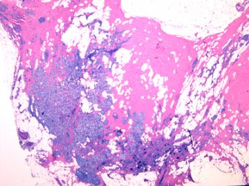
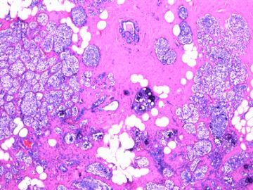
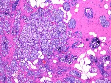
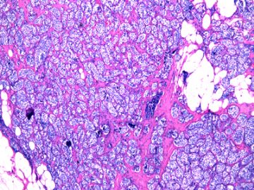
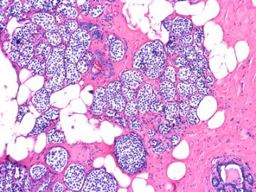
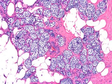
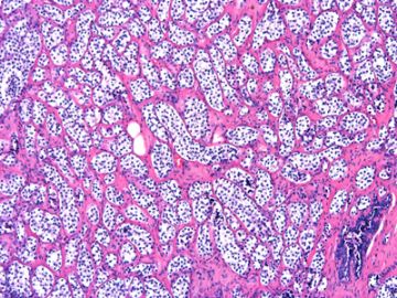
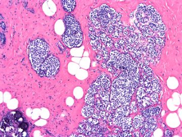
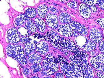
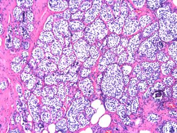
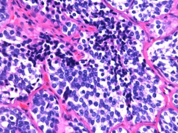
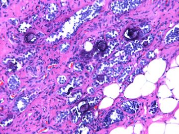
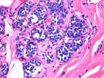
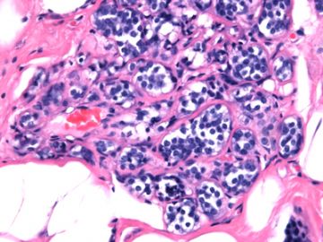
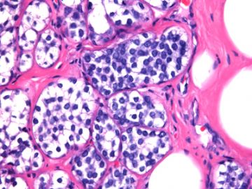
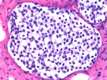
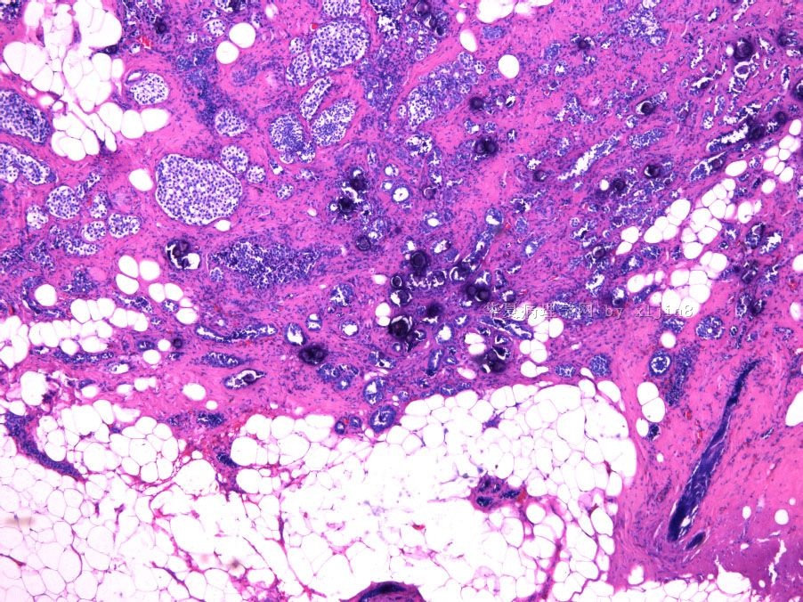
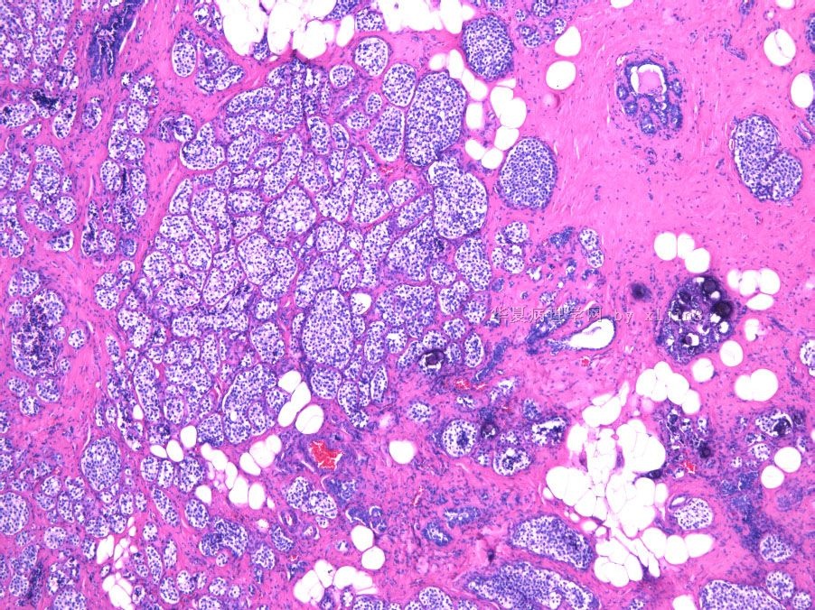
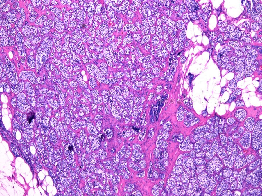
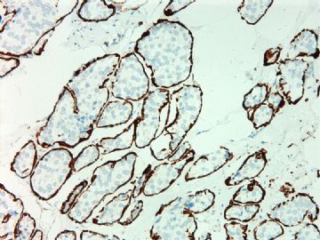
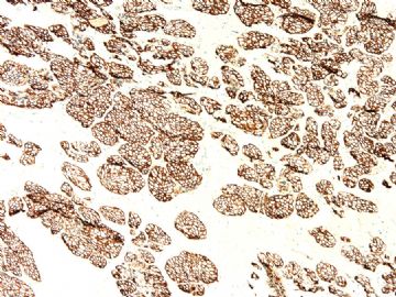
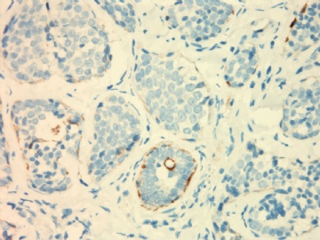
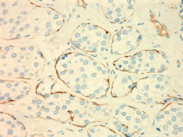
- B2694女性/52岁 右乳腺肿块伴钙化
| 姓 名: | ××× | 性别: | 年龄: | ||
| 标本名称: | 右乳腺病变切除标本 | ||||
| 简要病史: | 钼靶示右乳腺不规则钙化一年半,近一月来触及肿块0.5cm。 | ||||
| 肉眼检查: | 组织一块,5.5x3.5x2.5cm,切开有砂砾感,灰红色与灰白色相间,结节不明显。 | ||||
标签:DCIS LCIS

- xljin8
相关帖子
- • 左乳腺肿物
- • 乳腺癌?
- • 乳腺肿物
- • 乳腺肿物
- • 左乳癌标本乳头一个导管内的病变
- • 乳腺两个相邻导管内的病变
- • 乳腺肿物,请各位老师帮忙会诊
- • 女 46岁发现左乳腺肿块一月余
- • 乳腺肿物
- • 乳腺肿物
×参考诊断
Very extensive LCIS, should look hard for invasive lobular or ductal carcinoma, especially in sclerosing area.
Lobular neoplasm (atypical lobular hyperplasia and LCIS) is associated with increased risk for breast cancer of all kinds including ductal carcinoma, even the contralateral breast.
-
本帖最后由 于 2010-06-05 12:12:00 编辑
LCIS and ALH involving sclerosing adenosis and with microcalcifications.
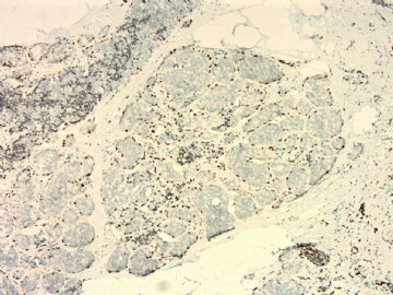
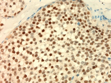
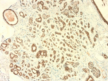
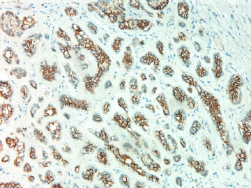
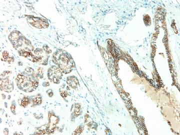
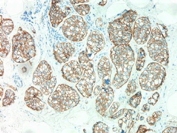
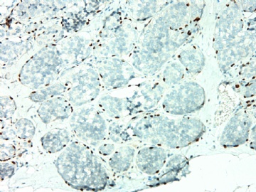
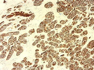
Currently we stain for almost all lobular lesions (e-cad/p120).
May stain myoepithelial markers if invasion cannot be excluded by H&E. I feel there is no invasion component.
(abin译:小叶原位癌LCIS和小叶不典型增生累及硬化性腺病,并有钙化。
目前我们几乎对所有小叶病变作免疫染色(e-cad/p120)。
如果HE不能排除浸润,也可染肌上皮标记物。我觉得无浸润成分。)

