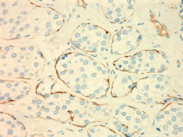
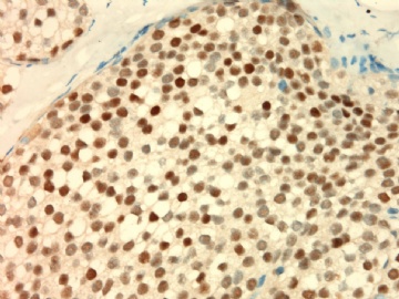
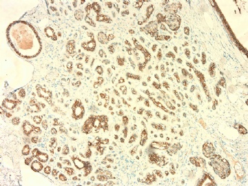
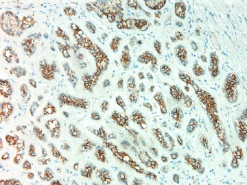
| 图片: | |
|---|---|
| 名称: | |
| 描述: | |
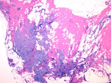
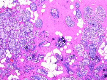
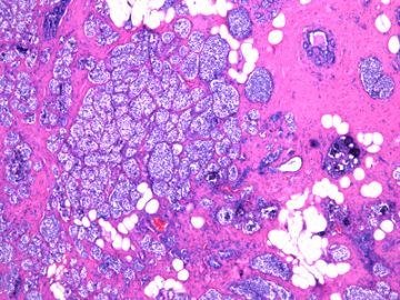
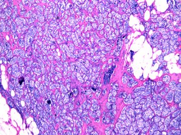
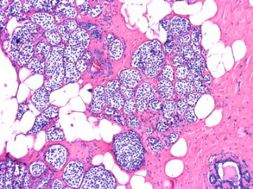
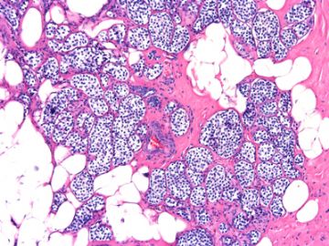
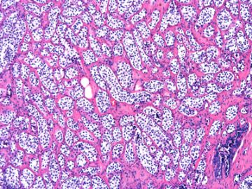
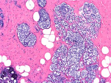
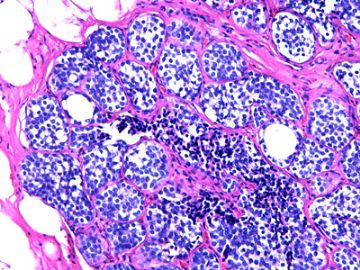
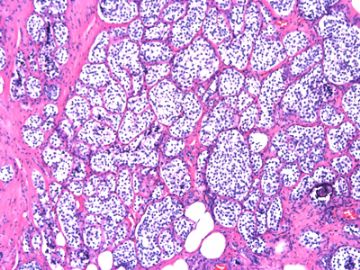
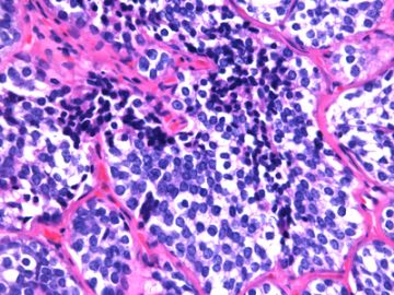
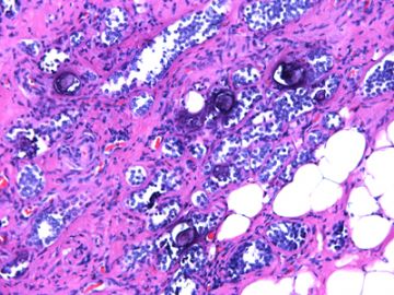
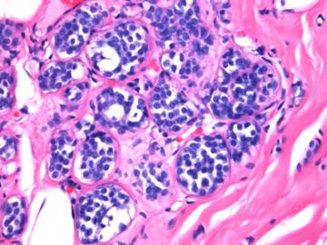
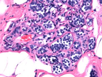
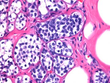
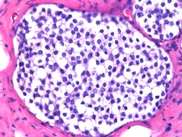
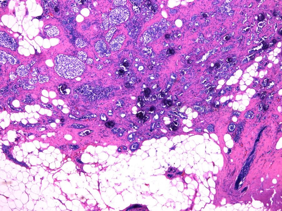
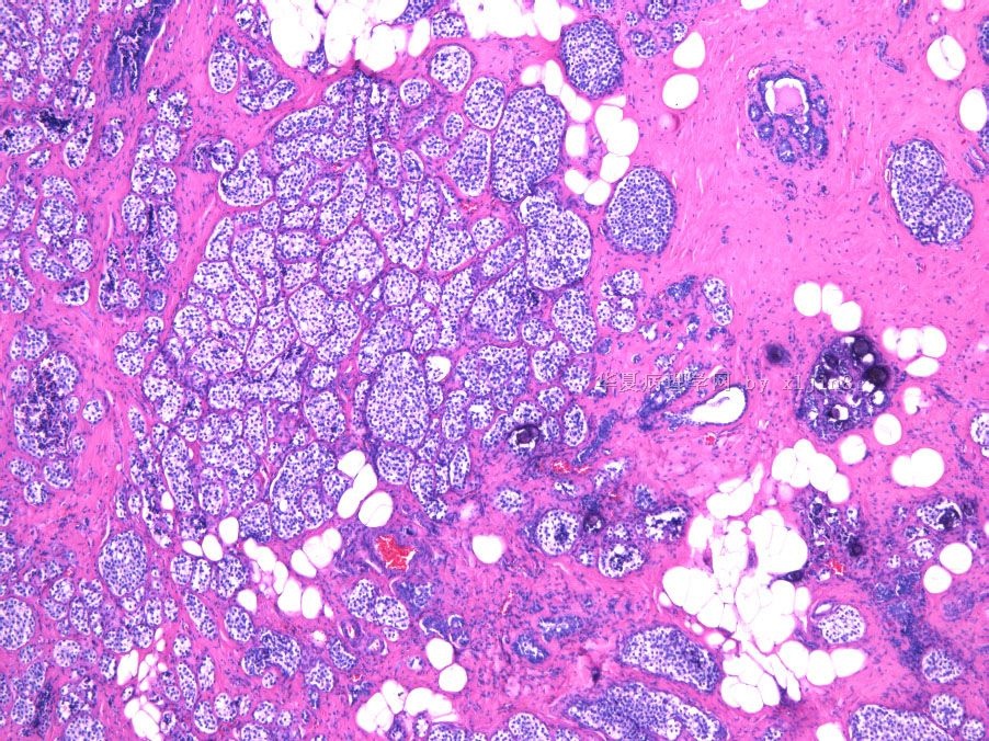
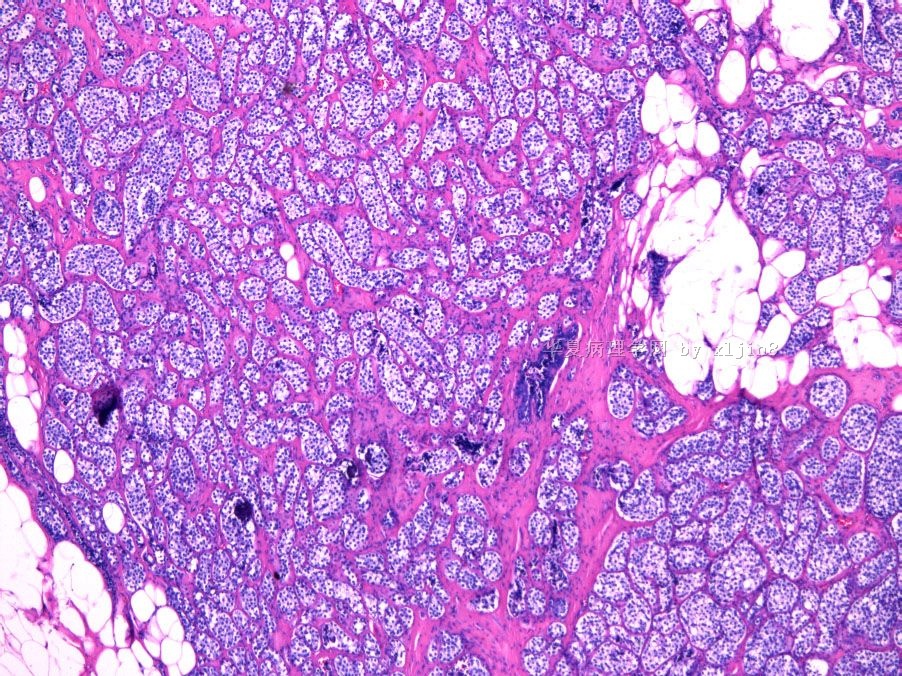
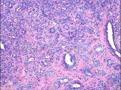
- B2694女性/52岁 右乳腺肿块伴钙化
| 姓 名: | ××× | 性别: | 年龄: | ||
| 标本名称: | 右乳腺病变切除标本 | ||||
| 简要病史: | 钼靶示右乳腺不规则钙化一年半,近一月来触及肿块0.5cm。 | ||||
| 肉眼检查: | 组织一块,5.5x3.5x2.5cm,切开有砂砾感,灰红色与灰白色相间,结节不明显。 | ||||

- xljin8
相关帖子
- • 左乳腺肿物
- • 乳腺癌?
- • 乳腺肿物
- • 乳腺肿物
- • 左乳癌标本乳头一个导管内的病变
- • 乳腺两个相邻导管内的病变
- • 乳腺肿物,请各位老师帮忙会诊
- • 女 46岁发现左乳腺肿块一月余
- • 乳腺肿物
- • 乳腺肿物
-
本帖最后由 于 2010-06-05 12:12:00 编辑
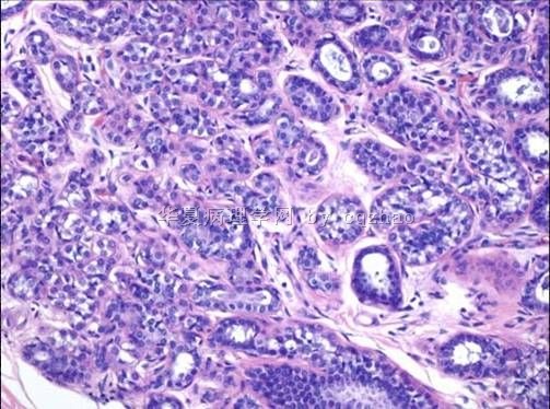
LCIS and ALH involving sclerosing adenosis and with microcalcifications.
Currently we stain for almost all lobular lesions (e-cad/p120).
May stain myoepithelial markers if invasion cannot be excluded by H&E. I feel there is no invasion component.
(abin译:小叶原位癌LCIS和小叶不典型增生累及硬化性腺病,并有钙化。
目前我们几乎对所有小叶病变作免疫染色(e-cad/p120)。
如果HE不能排除浸润,也可染肌上皮标记物。我觉得无浸润成分。)
Very extensive LCIS, should look hard for invasive lobular or ductal carcinoma, especially in sclerosing area.
Lobular neoplasm (atypical lobular hyperplasia and LCIS) is associated with increased risk for breast cancer of all kinds including ductal carcinoma, even the contralateral breast.
尝试回答金老师的提问:
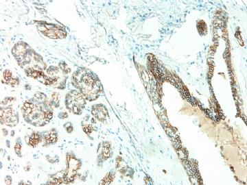
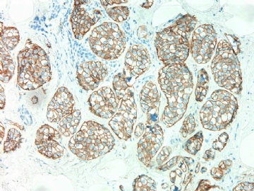
1)就IHC标记E-cadherin在导管-小叶单位有表达,是否为常见现象?
答:E-cadherin在正常导管-小叶单位的免疫表达,是常见现象。
2)在乳腺病的基础上,末端导管能否发生导管上皮化生,异表达E-cadherin?
答:腺病的“导管上皮化生”是否表示导管上皮发生各种化生性改变(鳞化、大汗腺化生)?所我们的观察,它们仍表达E-cadherin。
3) 此病例能否理解为高分化导管癌累及小叶(小叶癌化)?
可理解为低级别(或高分化)导管原位癌累及小叶(小叶癌化),不过小叶已经有硬化性腺病的基础病变。因此也可理解为低级别(或高分化)导管原位癌累及硬化性腺病。

华夏病理/粉蓝医疗
为基层医院病理科提供全面解决方案,
努力让人人享有便捷准确可靠的病理诊断服务。
-
本帖最后由 于 2010-06-08 21:17:00 编辑
Morphology is classic ALH or LCIS involving sclerosing adenosis. It is not like DCIS involving lobules.
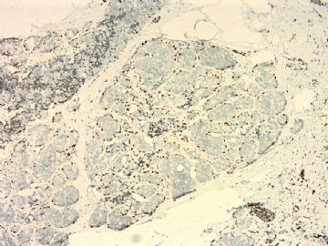
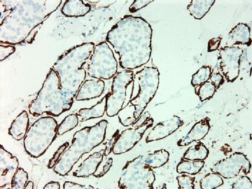
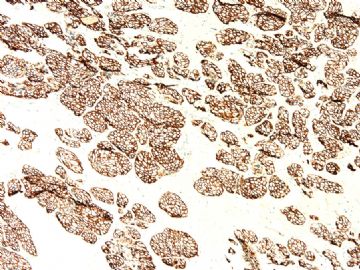
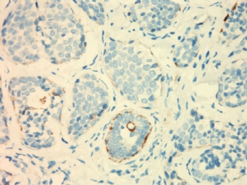
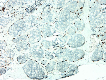
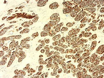
Now i tis the question how to evaluate the stain of E-cad for this case. Lobular lesions can show total loss or reduced expression of e-cad. Now you need to read yyour e-cad slides carefully and compare these sclerosing adenosis areas to normal ducts to determin if focal reduced expression of e-cad is present.
(abin译:形态学是典型的ALH或LCIS累及硬化性腺病。不像DCIS累及小叶。
现在我说说如何评估此例E-Ca染色。小叶病变可显示E-Ca表达的完全丢失或减少。这就需要仔细判读E-Ca染色切片,比较这些硬化性腺病区域与正常导管的显色差别,确定是否存在E-Ca表达的减少。)
-
本帖最后由 于 2010-06-08 21:20:00 编辑
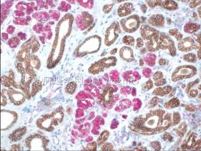
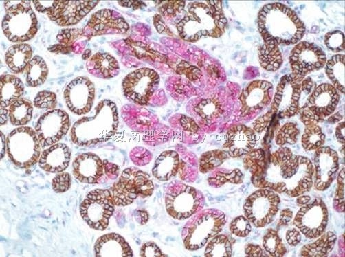
I have two cases sclerosing adenosis with focal ALH involvment. I used dual stains for these two cases. E-cad with bown color, p120 with red color. Clearly both cases show focal involvment with ALH.
(我提供两例硬化性腺病伴局灶ALH累及,使用了免疫双标。E-cad呈褐色,p120呈红色。两例均有明显的ALH局灶累及。)
| 以下是引用cqzhao在2010-6-8 5:24:00的发言:
Dr. Jin, I do not mean your case is certainly loblar lesion.I noticed that clear membrane stain of e-cad. But at least you can try to see if presence of e-cadexpression reduction. |
Dr. Jin,
我并不是说你的病例就是小叶病变。我注意到e-cad呈明确的膜着色。但至少可以努力观察是否存在e-cad表达减少。

华夏病理/粉蓝医疗
为基层医院病理科提供全面解决方案,
努力让人人享有便捷准确可靠的病理诊断服务。

