| 图片: | |
|---|---|
| 名称: | |
| 描述: | |
- 少见的肾脏肿块(新加免疫组化公布)
| 姓 名: | ××× | 性别: | 男 | 年龄: | 65 |
| 标本名称: | 左肾脏 | ||||
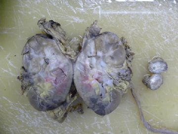
| 简要病史: | 肉眼血尿两天,影像学提示左肾脏占位。手术切除肾脏,发现腹主动脉旁有核桃大肿块,临床怀疑为肿大淋巴结。 | ||||
| 肉眼检查: | 左肾脏肿大,切面以肾盂为中心有4X4cm不规则灰白区,质硬,与正常肾组织无明显界限。腹主动脉旁肿块切面同肾脏。取材:1.肾被膜处;2.病变与正常组织交界处;3.灰白病变处;4.腹主动脉旁肿块。 | ||||
应金老师的提议,我们加做了HMB45,CD10,遗憾的是我科室没有Calponin。结果如图,请各位老师点评。
标签:
-
本帖最后由 于 2010-06-05 16:27:00 编辑

- 许春雷
×参考诊断
集合管癌
-
本帖最后由 于 2010-06-02 20:36:00 编辑
1)p63+ -- 集合管癌?
2)肌上皮癌?转移性或原发性?
3) IHC再标记:HMB-45、Calponin、CD10 WT-1
4)参考 Am J Surg Pathol 最新文献,少数集合管癌 p63可阳性(14%)
Albadine R, Schultz L, Illei P, Ertoy D, Hicks J, Sharma R, Epstein JI, Netto GJ.
PAX8 (+)/p63 (-) Immunostaining Pattern in Renal Collecting Duct Carcinoma (CDC):
A Useful Immunoprofile in the Differential Diagnosis of CDC Versus UrothelialCarcinoma of Upper Urinary Tract. Am J Surg Pathol. 2010 May 11. [Epub ahead of print]Departments of *Pathology double daggerUrology section signOncology, JohnsHopkins University, Baltimore, MD daggerDepartment of Oncology, HacettepeUniversity, Ankara, Turkey.
rare but aggressive type of renal malignancy with variable morphologic features.
One of the World Health Organization diagnostic criteria for CDC is the exclusion of
urothelial carcinoma of renal pelvis from the differential diagnosis. PAX8 is a novellineage restricted transcription factor expressed in renal tubules. We investigated the
expression pattern of PAX8 in CDC and its utility, in combination with p63, in resolving
the differential diagnosis of CDC versus upper tract urothelial carcinoma (UUC).
DESIGN: Archival tissues from 21 CDC and 34 UUC were retrieved from our institutional
files. Immunohistochemistry for PAX8 and p63 were performed on routine and tissue
microarray sections using standard immunohistochemistry protocol. Intensity of nuclear
staining was evaluated for each marker and assigned an incremental 0, 1+, 2+, and 3+ score. Extent
of staining was categorized as focal (<25%), nonfocal (25% to 75%), or diffuse (>75%).
RESULTS: CDC: All 21 (100%) CDC were positive for PAX8. Intensity of expression
was moderate to strong (2+/3+) in 19 cases (90%). Extent of staining was diffuse
in 13 of 21 tumors. The p63 was positive in 3 of 21 (14%) CDC cases (PAX8+/p63+).
UUC: The 34 UUC included 5 pT1, 4 pT2, and 25 pT3/pT4 tumors.Thirty-one of 34 (91.2%)
UUC were negative for PAX8, whereas 33 of 34 (97%) were p63 positive. Staining
intensity was moderate in 15 cases (44%), of which 12 were nonfocal or diffuse.
The unique p63-negative UUC was a pT1 tumor that was also negative for PAX8 (PAX8-/p63-).
CONCLUSIONS: We propose the use of the combination of PAX8 and p63 in the diagnosis of poorly
differentiated renal sinus epithelial neoplasms where the differential diagnosis includes
CDC versus UUC.The immunoprofile of PAX8+/p63- supports the diagnosis of CDC with a sensitivity
of 85.7% and a specificity of 100%. In contrast, a (PAX8-/p63+) profile supports the diagnosis
of UUC with a sensitivity of 88.2% and a specificity of 100%. The inverse PAX8/p63 expression
seen in CDC and UUC supports a renal tubular rather than an urothelial differentiation in CDC
given the nephric lineage restriction f PAX8.
- xljin8


