


| 图片: | |
|---|---|
| 名称: | |
| 描述: | |
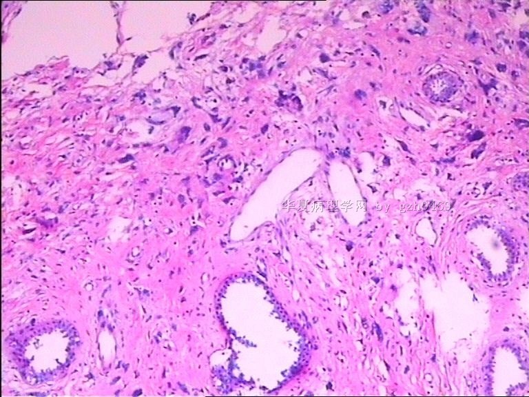
- 左乳肿物
-
本帖最后由 于 2010-05-28 12:07:00 编辑
Need to check carefully to see if there are mitoses. If you cannot see any mitosis, it is more like a fibroadennoma with multinucleated stromal giant cells or pleomorphic stromal giant cells.
It is benign.
abin译:需要仔细检查是否有核分裂。如果没有核分裂,很可能是纤维腺瘤伴多核间质巨细胞或多形性间质巨细胞。属于良性。
-
本帖最后由 于 2010-05-28 12:04:00 编辑
Benign tumors of the breast with multinucleated stromal giant cells. Immunohistochemical analysis of six cases and review of the literature.
Ryska A, Reynolds C, Keeney GL.
Department of Pathology, Charles University Medical Faculty Hospital, Hradec Králové, Czech Republic. ryskaale@fnhk.cz
Abstract
The authors present six cases of benign tumors of the breast with numerous multinucleated stromal giant cells (MSGC). All six patients were women aged 37-70 years (mean 48 years), presenting clinically with a breast mass 1.0-3.8 cm in size (mean 1.9 cm; median 1.5 cm). By standard H&E examination, all cases showed the presence of numerous MSGC haphazardly dispersed within the tumor stroma. Three cases revealed MSGC merging into the surrounding adipose tissue simulating infiltrative growth. The MSGC appeared to have multiple nuclei (5 to 25) with fine chromatin and sporadic small nucleoli. Their cytoplasm was inconspicuous. The MSGC expressed vimentin only and to lesser extent CD34. These cells were negative for muscle markers, keratins, S-100 protein, vascular markers, CD68 and hormone receptors. Interestingly, the majority of MSGC and mononuclear stromal cells showed reactivity for p53 protein and Ki-67 proliferation antigen. All patients were treated by simple excision and remain free of recurrence (mean 70 months, median 48 months.). The reactivity of p53 in MSGC and mononuclear stromal cells may play a key role in linking these two cell types. Nonetheless, the presence of MSGC does not alter prognosis of otherwise typical benign lesions.
-
本帖最后由 于 2010-05-28 12:04:00 编辑
Ann Diagn Pathol. 2009 Aug;13(4):226-32. Epub 2009 May 23.
Fibroepithelial lesions of the breast with pleomorphic stromal giant cells: a clinicopathologic study of 4 cases and review of the literature.
Department of Pathology and Laboratory Medicine, The University of Texas, MD Anderson Cancer Center, Houston, TX 77030, USA. leihuo@mdanderson.org
Abstract
Pleomorphic stromal giant cells are occasionally found as an incidental finding in breast tissue but are only rarely seen in fibroepithelial lesions. In this report, we describe 4 fibroadenoma-like lesions of the breast with pleomorphic stromal giant cells. Two cases had focal stromal hypercellularity, one of which was with architectural features borderline between a fibroadenoma and a phyllodes tumor, but none was considered diagnostic of phyllodes tumor. One lesion had up to 4 mitotic figures per 10 high-power fields, including rare atypical mitotic figures. The remaining 3 cases lacked mitotic activity. Follow-up for 3 cases at 16 to 59 months revealed no evidence of tumor recurrence. The fourth case was lost to follow-up. It appears that the presence of pleomorphic stromal giant cells in an otherwise benign fibroepithelial lesion has no adverse clinical significance. The clinicopathologic features of each case are discussed, and a review of the literature is provided
-
本帖最后由 于 2010-05-28 12:05:00 编辑
Diagn Pathol. 2008 Aug 1;3:33.
A diagnostic dilemma in breast pathology--benign fibroadenoma with multinucleated stromal giant cells.
Heneghan HM, Martin ST, Casey M, Tobbia I, Benani F, Barry KM.
Department of Surgery, Mayo General Hospital, Ireland. helenheneghan@hotmail.com
Abstract
Fibroadenomas are common benign breast tumours that display a characteristic pathological morphology, although several epithelial and stromal variations exist. A very rare histological finding is the presence of multinucleated giant cells throughout the stroma of a benign fibroadenoma. Cells of this type, which are more commonly found incidentally within the interlobular stroma of breast tissue, are benign and should not be mistaken for malignant cells on microscopic examination. Unfortunately a lack of awareness of this pathological entity can lead to diagnostic confusion amongst pathologists resulting in the multinucleate giant cells being mistaken for highly mitotic cells and consequently the fibroadenoma being mistaken for a malignant lesion. This may have serious implications for the subsequent management of the patient. The presence of this unusual cell type in the stroma does not alter the prognosis of otherwise benign lesion. We encountered two such cases at our institution in a six month period recently. We present their histories along with relevant radiological, microscopic and immunohistochemical features, followed by a discussion of this unusual pathological entity.
Dr.cqzhao提供的文献,要点翻译如下:
3楼
免疫组化:多核巨细胞仅表达Vim,CD34表达较弱。阴性:肌源性标记物、角蛋白、S-100、血管标记物、CD68和激素受体。大多数多核巨细胞和单核间质细胞表达p53和Ki67。存在多核巨细胞不会改变其它方面呈典型良性病变的预后。
4楼
在其它方面表现为良性的纤维上皮性病变,出现多核巨细胞没有不利的临床意义。
5楼
如果认识不足,会误认为核分裂细胞,将纤维腺瘤误诊为恶性。对于其它方面良性的病例,存在多核巨细胞并不改变预后。

华夏病理/粉蓝医疗
为基层医院病理科提供全面解决方案,
努力让人人享有便捷准确可靠的病理诊断服务。


