| 图片: | |
|---|---|
| 名称: | |
| 描述: | |
- B2686乳腺包块
| 姓 名: | ××× | 性别: | 女 | 年龄: | 20 |
| 标本名称: | 纤维腺瘤 | ||||
| 简要病史: | 发现乳腺包块2个月 | ||||
| 肉眼检查: | 结节状组织一块,大小1.5*1*0.8,切实,切面灰白,质中 | ||||

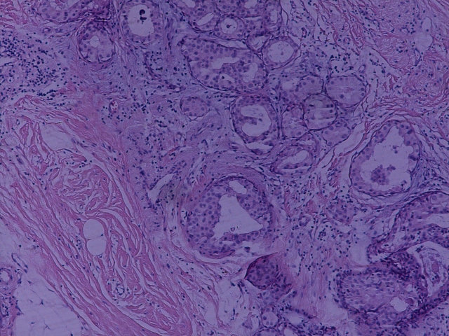
名称:图1
描述:图1

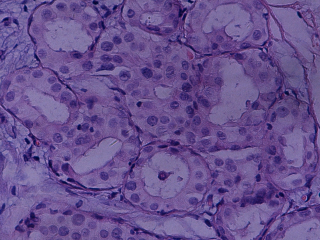
名称:图2
描述:图2

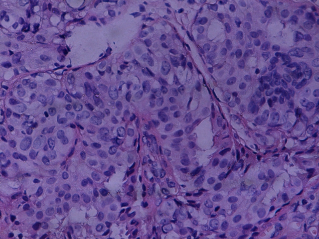
名称:图3
描述:图3

- 典型中看不典型,不典型中找典型。
相关帖子
- • 左乳腺肿物
- • 乳腺癌?
- • 乳腺肿物
- • 乳腺肿物
- • 左乳癌标本乳头一个导管内的病变
- • 乳腺两个相邻导管内的病变
- • 乳腺肿物,请各位老师帮忙会诊
- • 女 46岁发现左乳腺肿块一月余
- • 乳腺肿物
- • 乳腺肿物
-
CHENYINQIAO 离线
- 帖子:547
- 粉蓝豆:0
- 经验:593
- 注册时间:2010-07-20
- 加关注 | 发消息
| 以下是引用晓明在2010-5-29 0:44:00的发言:
不好意思,导管周围肌上皮存在且连续,导管内细胞增生单一,大部分是导管上皮超过90%。 E-cadherin阳性,肌上皮标记我们一般用P63,SMA,calponin和CK34E12 等我找一下免疫组化传一下 |
E-cad positive excludes lobular lesion. Myoepithelial stains are no meaning in this case. The key point for this case is the lesion is DCIS or ADH. Except for this young women has DCIS in other area, the photos may represent DCIS with lobular involvment. Other wise I feel it is better to call ADH.
If the specimen is breast core biopsy the patient need to have excisonal biopsy it does not matter what we call dcis or adh.
If this is a excisional biopsy with clear margins, our pathology report will be very important for next clinical management of the young lady.
If we call ADH, we do not need to report the margin and the women will have close clinical and imagingfollow up. If we call DCIS, pathologists need to report the size of DCIS, margins, do ER, PR stains. Breasy surgeons or oncologists need to consider the possibility of sentinel lymph node biopsy, radiation, adjuvant chemotherapy or total mastectomy based on individual patients.
So pathology diagnnosis guides the clincial managment. This is why we must be very cautious to make our dx.


 能否上传免疫组化图片?谢谢


